3GSQ | pdb_00003gsq

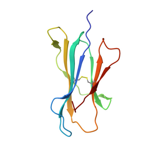
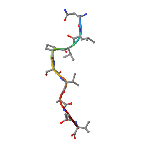
Crystal structure of the binary complex between HLA-A2 and HCMV NLV-M5S peptide variant


Experimental Data Snapshot

  • Method: X-RAY DIFFRACTION
  • Resolution: 2.12 Å
  • R-Value Free: 
    0.246 (Depositor) 
  • R-Value Work: 
    0.196 (Depositor) 
  • R-Value Observed: 
    0.199 (Depositor) 

wwPDB Validation 3D Report Full Report

Validation slider image for 3GSQ

This is version 1.3 of the entry. See complete history. 

Literature

Structural bases for the affinity-driven selection of a public TCR against a dominant human cytomegalovirus epitope.

Gras, S., Saulquin, X., Reiser, J.B., Debeaupuis, E., Echasserieau, K., Kissenpfennig, A., Legoux, F., Chouquet, A., Le Gorrec, M., Machillot, P., Neveu, B., Thielens, N., Malissen, B., Bonneville, M., Housset, D.

(2009) J Immunol 183: 430-437

  • DOI: https://doi.org/10.4049/jimmunol.0900556
  • Primary Citation Related Structures: 
    3GSN, 3GSO, 3GSQ, 3GSR, 3GSU, 3GSV, 3GSW, 3GSX

  • PubMed Abstract: 

    Protective T cell responses elicited along chronic human CMV (HCMV) infections are sometimes dominated by CD8 T cell clones bearing highly related or identical public TCR in unrelated individuals. To understand the principles that guide emergence of these public T cell responses, we have performed structural, biophysical, and functional analyses of an immunodominant public TCR (RA14) directed against a major HLA-A*0201-restricted HCMV Ag (pp65(495-503)) and selected in vivo from a diverse repertoire after chronic stimulations. Unlike the two immunodominant public TCRs crystallized so far, which focused on one peptide hotspot, the HCMV-specific RA14 TCR interacts with the full array of available peptide residues. The conservation of some peptide-MHC complex-contacting amino acids by lower-affinity TCRs suggests a shared TCR-peptide-MHC complex docking mode and supports an Ag-driven selection of optimal TCRs. Therefore, the emergence of a public TCR of an oligoclonal Ag-specific response after repeated viral stimulations is based on a receptor displaying a high structural complementarity with the entire peptide and focusing on three peptide hotspots. This highlights key parameters underlying the selection of a protective T cell response against HCMV infection, which remains a major health issue in patients undergoing bone marrow transplantation.


  • Organizational Affiliation: 
    • Institut de Biologie Structurale Jean-Pierre Ebel, CEA, CNRS, UJF, PSB, Grenoble, France.

Macromolecule Content 

  • Total Structure Weight: 44.53 kDa 
  • Atom Count: 3,370 
  • Modeled Residue Count: 383 
  • Deposited Residue Count: 383 
  • Unique protein chains: 3

Macromolecules

Find similar proteins by:|  3D Structure
Entity ID: 1
MoleculeChains  Sequence LengthOrganismDetailsImage
HLA class I histocompatibility antigen, A-2 alpha chain274Homo sapiensMutation(s): 1 
Gene Names: HLA, HLA-A, HLAA
UniProt & NIH Common Fund Data Resources
Find proteins for P04439 (Homo sapiens)
Explore P04439 
Go to UniProtKB:  P04439
PHAROS:  P04439
GTEx:  ENSG00000206503 
Entity Groups
Sequence Clusters30% Identity50% Identity70% Identity90% Identity95% Identity100% Identity
UniProt GroupP04439
Sequence Annotations
Expand
Reference Sequence
Find similar proteins by:|  3D Structure
Entity ID: 2
MoleculeChains  Sequence LengthOrganismDetailsImage
Beta-2-microglobulin100Homo sapiensMutation(s): 0 
Gene Names: B2M, Beta-2 microglubulin, CDABP0092, HDCMA22P
UniProt & NIH Common Fund Data Resources
Find proteins for P61769 (Homo sapiens)
Explore P61769 
Go to UniProtKB:  P61769
PHAROS:  P61769
GTEx:  ENSG00000166710 
Entity Groups
Sequence Clusters30% Identity50% Identity70% Identity90% Identity95% Identity100% Identity
UniProt GroupP61769
Sequence Annotations
Expand
Reference Sequence
Find similar proteins by:  Sequence   |   3D Structure  
Entity ID: 3
MoleculeChains  Sequence LengthOrganismDetailsImage
HCMV pp65 fragment 495-503, variant M5S (NLVPSVATV)C [auth P]9N/AMutation(s): 0 
Sequence Annotations
Expand
Reference Sequence

Experimental Data & Validation

Experimental Data

  • Method: X-RAY DIFFRACTION
  • Resolution: 2.12 Å
  • R-Value Free:  0.246 (Depositor) 
  • R-Value Work:  0.196 (Depositor) 
  • R-Value Observed: 0.199 (Depositor) 
Space Group: P 1 21 1
Unit Cell:
Length ( Å )Angle ( ˚ )
a = 54.047α = 90
b = 80.897β = 114.17
c = 57.659γ = 90
Software Package:
Software NamePurpose
XSCALEdata scaling
AMoREphasing
REFMACrefinement
PDB_EXTRACTdata extraction
ADSCdata collection
XDSdata reduction

Structure Validation

View Full Validation Report


Entry History 

Revision History  (Full details and data files)

  • Version 1.0: 2009-08-04
    Type: Initial release
  • Version 1.1: 2011-07-13
    Changes: Version format compliance
  • Version 1.2: 2021-10-20
    Changes: Database references
  • Version 1.3: 2024-11-06
    Changes: Data collection, Structure summary