3GOX | pdb_00003gox

Crystal structure of the beta-beta-alpha-Me type II restriction endonuclease Hpy99I in the absence of EDTA


Experimental Data Snapshot

  • Method: X-RAY DIFFRACTION
  • Resolution: 1.50 Å
  • R-Value Free: 
    0.186 (Depositor), 0.211 (DCC) 
  • R-Value Work: 
    0.169 (Depositor), 0.193 (DCC) 
  • R-Value Observed: 
    0.170 (Depositor) 

wwPDB Validation 3D Report Full Report

Validation slider image for 3GOX

This is version 1.3 of the entry. See complete history

Literature

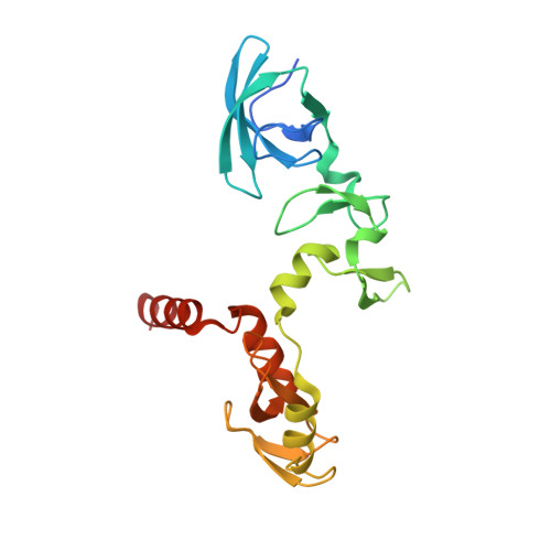
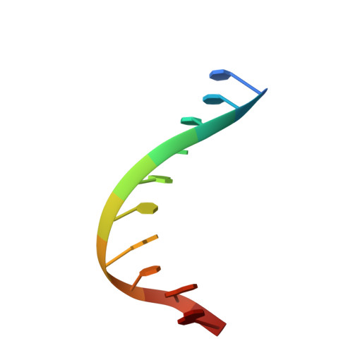
Crystal structure of the beta beta alpha-Me type II restriction endonuclease Hpy99I with target DNA.

Sokolowska, M.Czapinska, H.Bochtler, M.

(2009) Nucleic Acids Res 37: 3799-3810

  • DOI: https://doi.org/10.1093/nar/gkp228
  • Primary Citation Related Structures: 
    3FC3, 3GOX

  • PubMed Abstract: 

    The beta beta alpha-Me restriction endonuclease (REase) Hpy99I recognizes the CGWCG target sequence and cleaves it with unusual stagger (five nucleotide 5'-recessed ends). Here we present the crystal structure of the specific complex of the dimeric enzyme with DNA. The Hpy99I protomer consists of an antiparallel beta-barrel and two beta 4 alpha 2 repeats. Each repeat coordinates a structural zinc ion with four cysteine thiolates in two CXXC motifs. The beta beta alpha-Me region of the second beta 4 alpha 2 repeat holds the catalytic metal ion (or its sodium surrogate) via Asp148 and Asn165 and activates a water molecule with the general base His149. In the specific complex, Hpy99I forms a ring-like structure around the DNA that contacts DNA bases on the major and minor groove sides via the first and second beta 4 alpha 2 repeats, respectively. Hpy99I interacts with the central base pair of the recognition sequence only on the minor groove side, where A:T resembles T:A and G:C is similar to C:G. The Hpy99I-DNA co-crystal structure provides the first detailed illustration of the beta beta alpha-Me site in REases and complements structural information on the use of this active site motif in other groups of endonucleases such as homing endonucleases (e.g. I-PpoI) and Holliday junction resolvases (e.g. T4 endonuclease VII).


  • Organizational Affiliation
    • International Institute of Molecular and Cell Biology, Trojdena 4, 02-109 Warsaw, Poland.

Macromolecule Content 

  • Total Structure Weight: 52.81 kDa 
  • Atom Count: 4,411 
  • Modeled Residue Count: 398 
  • Deposited Residue Count: 422 
  • Unique protein chains: 1
  • Unique nucleic acid chains: 2

Macromolecules


Find similar proteins by:|  3D Structure
Entity ID: 1
MoleculeChains  Sequence LengthOrganismDetailsImage
Restriction endonuclease Hpy99I
A, B
200Helicobacter pylori J99Mutation(s): 0 
Gene Names: jhp_0755synthetic gene codon optimized for Escherichia coli
UniProt
Find proteins for Q9ZL26 (Helicobacter pylori (strain J99 / ATCC 700824))
Explore Q9ZL26 
Go to UniProtKB:  Q9ZL26
Entity Groups
Sequence Clusters30% Identity50% Identity70% Identity90% Identity95% Identity100% Identity
UniProt GroupQ9ZL26
Sequence Annotations
Expand
Reference Sequence
Find similar nucleic acids by:  Sequence
Entity ID: 2
MoleculeChains LengthOrganismImage
5'-(*DCP*DTP*DCP*DGP*DAP*DCP*DGP*DTP*DAP*DGP*DA)-3'11N/A
Sequence Annotations
Expand
Reference Sequence
Find similar nucleic acids by:  Sequence
Entity ID: 3
MoleculeChains LengthOrganismImage
5'-(*DTP*DAP*DCP*DGP*DTP*DCP*DGP*DAP*DGP*DTP*DC)-3'11N/A
Sequence Annotations
Expand
Reference Sequence

Small Molecules

Ligands 3 Unique
IDChains Name / Formula / InChI Key2D Diagram3D Interactions
1PE

Query on 1PE


Download:Ideal Coordinates CCD File
H [auth A]PENTAETHYLENE GLYCOL
C10 H22 O6
JLFNLZLINWHATN-UHFFFAOYSA-N
ZN

Query on ZN


Download:Ideal Coordinates CCD File
E [auth A],
F [auth A],
I [auth B],
J [auth B]
ZINC ION
Zn
PTFCDOFLOPIGGS-UHFFFAOYSA-N
NA

Query on NA


Download:Ideal Coordinates CCD File
G [auth A],
K [auth B],
L [auth B]
SODIUM ION
Na
FKNQFGJONOIPTF-UHFFFAOYSA-N

Experimental Data & Validation

Experimental Data

  • Method: X-RAY DIFFRACTION
  • Resolution: 1.50 Å
  • R-Value Free:  0.186 (Depositor), 0.211 (DCC) 
  • R-Value Work:  0.169 (Depositor), 0.193 (DCC) 
  • R-Value Observed: 0.170 (Depositor) 
Space Group: H 3 2
Unit Cell:
Length ( Å )Angle ( ˚ )
a = 90.682α = 90
b = 90.682β = 90
c = 334.933γ = 120
Software Package:
Software NamePurpose
MxCuBEdata collection
SHELXCDphasing
SHELXDphasing
SHELXEmodel building
ARP/wARPmodel building
REFMACrefinement
XDSdata reduction
XSCALEdata scaling

Structure Validation

View Full Validation Report


Entry History 

Deposition Data

Revision History  (Full details and data files)

  • Version 1.0: 2009-04-28
    Type: Initial release
  • Version 1.1: 2011-07-13
    Changes: Source and taxonomy, Version format compliance
  • Version 1.2: 2017-11-01
    Changes: Advisory, Refinement description
  • Version 1.3: 2024-02-21
    Changes: Advisory, Data collection, Database references, Derived calculations