The juxtamembrane region of the EGF receptor functions as an activation domain.
Red Brewer, M., Choi, S.H., Alvarado, D., Moravcevic, K., Pozzi, A., Lemmon, M.A., Carpenter, G.(2009) Mol Cell 34: 641-651
- PubMed: 19560417 Search on PubMedSearch on PubMed Central
- DOI: https://doi.org/10.1016/j.molcel.2009.04.034
- Primary Citation Related Structures:
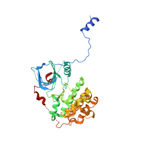
3GOP - PubMed Abstract:
In several growth factor receptors, the intracellular juxtamembrane (JM) region participates in autoinhibitory interactions that must be disrupted for tyrosine kinase activation. Using alanine scanning mutagenesis and crystallographic approaches, we define a domain within the JM region of the epidermal growth factor receptor (EGFR) that instead plays an activating--rather than autoinhibitory--role. Mutations in the C-terminal 19 residues of the EGFR JM region abolish EGFR activation. In a crystal structure of an asymmetric dimer of the tyrosine kinase domain, the JM region of an acceptor monomer makes extensive contacts with the C lobe of a donor monomer, thus stabilizing the dimer. We describe how an uncharacterized lung cancer mutation in this JM activation domain (V665M) constitutively activates EGFR by augmenting its capacity to act as an acceptor in the asymmetric dimer. This JM mutant promotes cellular transformation by EGFR in vitro and is tumorigenic in a xenograft assay.
- Department of Biochemistry, Vanderbilt University School of Medicine, Nashville, TN 37212, USA.
Organizational Affiliation: