3GJQ

Caspase-3 Binds Diverse P4 Residues in Peptides


Experimental Data Snapshot

  • Method: X-RAY DIFFRACTION
  • Resolution: 2.60 Å
  • R-Value Free: 0.290 
  • R-Value Work: 0.236 

wwPDB Validation   3D Report Full Report


This is version 1.1 of the entry. See complete history


Literature

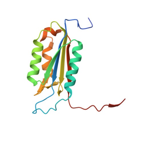
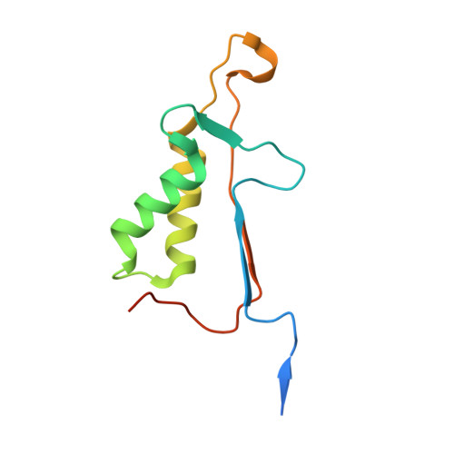
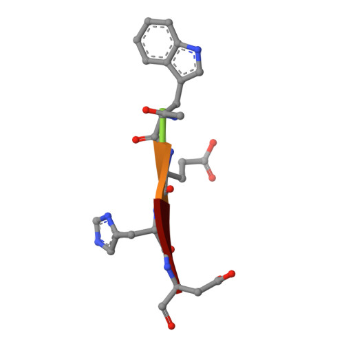
Caspase-3 binds diverse P4 residues in peptides as revealed by crystallography and structural modeling.

Fang, B.Fu, G.Agniswamy, J.Harrison, R.W.Weber, I.T.

(2009) Apoptosis 14: 741-752

  • DOI: https://doi.org/10.1007/s10495-009-0333-y
  • Primary Citation of Related Structures:  
    3GJQ, 3GJR, 3GJS, 3GJT

  • PubMed Abstract: 

    Caspase-3 recognition of various P4 residues in its numerous protein substrates was investigated by crystallography, kinetics, and calculations on model complexes. Asp is the most frequent P4 residue in peptide substrates, although a wide variety of P4 residues are found in the cellular proteins cleaved by caspase-3. The binding of peptidic inhibitors with hydrophobic P4 residues, or no P4 residue, is illustrated by crystal structures of caspase-3 complexes with Ac-IEPD-Cho, Ac-WEHD-Cho, Ac-YVAD-Cho, and Boc-D(OMe)-Fmk at resolutions of 1.9-2.6 A. The P4 residues formed favorable hydrophobic interactions in two separate hydrophobic regions of the binding site. The side chains of P4 Ile and Tyr form hydrophobic interactions with caspase-3 residues Trp206 and Trp214 within a non-polar pocket of the S4 subsite, while P4 Trp interacts with Phe250 and Phe252 that can also form the S5 subsite. These interactions of hydrophobic P4 residues are distinct from those for polar P4 Asp, which indicates the adaptability of caspase-3 for binding diverse P4 residues. The predicted trends in peptide binding from molecular models had high correlation with experimental values for peptide inhibitors. Analysis of structural models for the binding of 20 different amino acids at P4 in the aldehyde peptide Ac-XEVD-Cho suggested that the majority of hydrophilic P4 residues interact with Phe250, while hydrophobic residues interact with Trp206, Phe250, and Trp214. Overall, the S4 pocket of caspase-3 exhibits flexible adaptation for different residues and the new structures and models, especially for hydrophobic P4 residues, will be helpful for the design of caspase-3 based drugs.


  • Organizational Affiliation

    Department of Biology, Molecular Basis of Disease Program, Georgia State University, Atlanta, GA 30303, USA.


Macromolecules
Find similar proteins by:  (by identity cutoff)  |  3D Structure
Entity ID: 1
MoleculeChains Sequence LengthOrganismDetailsImage
Caspase-3 subunit p17
A, C
147Homo sapiensMutation(s): 0 
Gene Names: CASP3CPP32
EC: 3.4.22.56
UniProt & NIH Common Fund Data Resources
Find proteins for P42574 (Homo sapiens)
Explore P42574 
Go to UniProtKB:  P42574
PHAROS:  P42574
GTEx:  ENSG00000164305 
Entity Groups  
Sequence Clusters30% Identity50% Identity70% Identity90% Identity95% Identity100% Identity
UniProt GroupP42574
Sequence Annotations
Expand
  • Reference Sequence
Find similar proteins by:  (by identity cutoff)  |  3D Structure
Entity ID: 2
MoleculeChains Sequence LengthOrganismDetailsImage
Caspase-3 subunit p12
B, D
108Homo sapiensMutation(s): 0 
Gene Names: CASP3CPP32
EC: 3.4.22.56
UniProt & NIH Common Fund Data Resources
Find proteins for P42574 (Homo sapiens)
Explore P42574 
Go to UniProtKB:  P42574
PHAROS:  P42574
GTEx:  ENSG00000164305 
Entity Groups  
Sequence Clusters30% Identity50% Identity70% Identity90% Identity95% Identity100% Identity
UniProt GroupP42574
Sequence Annotations
Expand
  • Reference Sequence

Find similar proteins by:  Sequence   |   3D Structure  

Entity ID: 3
MoleculeChains Sequence LengthOrganismDetailsImage
peptide inhibitor
E, F
5N/AMutation(s): 0 
Sequence Annotations
Expand
  • Reference Sequence
Experimental Data & Validation

Experimental Data

  • Method: X-RAY DIFFRACTION
  • Resolution: 2.60 Å
  • R-Value Free: 0.290 
  • R-Value Work: 0.236 
  • Space Group: P 21 21 21
Unit Cell:
Length ( Å )Angle ( ˚ )
a = 69.099α = 90
b = 88.247β = 90
c = 96.634γ = 90
Software Package:
Software NamePurpose
HKL-2000data collection
AMoREphasing
CNSrefinement
HKL-2000data reduction
HKL-2000data scaling

Structure Validation

View Full Validation Report


Entry History 

Deposition Data

Revision History  (Full details and data files)

  • Version 1.0: 2009-03-24
    Type: Initial release
  • Version 1.1: 2011-07-13
    Changes: Version format compliance