3GIS

Crystal Structure of Na-free Thrombin in Complex with Thrombomodulin


Experimental Data Snapshot

  • Method: X-RAY DIFFRACTION
  • Resolution: 2.40 Å
  • R-Value Free: 0.259 
  • R-Value Work: 0.211 
  • R-Value Observed: 0.243 

Starting Models: experimental
View more details

wwPDB Validation   3D Report Full Report


This is version 1.3 of the entry. See complete history


Literature

Molecular basis of thrombomodulin activation of slow thrombin

Adams, T.E.Li, W.Huntington, J.A.

(2009) J Thromb Haemost 7: 1688-1695

  • DOI: https://doi.org/10.1111/j.1538-7836.2009.03563.x
  • Primary Citation of Related Structures:  
    3GIS

  • PubMed Abstract: 

    Coagulation is a highly regulated process where the ability to prevent blood loss after injury is balanced against the maintenance of blood fluidity. Thrombin is at the center of this balancing act. It is the critical enzyme for producing and stabilizing a clot, but when complexed with thrombomodulin (TM) it is converted to a powerful anticoagulant. Another cofactor that may play a role in determining thrombin function is the monovalent cation Na(+). Its apparent affinity suggests that half of the thrombin generated is in a Na(+)-free 'slow' state and half is in a Na(+)-coordinated 'fast' state. While slow thrombin is a poor procoagulant enzyme, when complexed to TM it is an effective anticoagulant. To better understand this molecular transformation we solved a 2.4 A structure of thrombin complexed with EGF domains 4-6 of TM in the absence of Na(+) and other cofactors or inhibitors. We find that TM binds as previously observed, and that the thrombin component resembles structures of the fast form. The Na(+) binding loop is observed in a conformation identical to the Na(+)-bound form, with conserved water molecules compensating for the missing ion. Using the fluorescent probe p-aminobenzamidine we show that activation of slow thrombin by TM principally involves the opening of the primary specificity pocket. These data show that TM binding alters the conformation of thrombin in a similar manner as Na(+) coordination, resulting in an ordering of the Na(+) binding loop and an opening of the adjacent S1 pocket. We conclude that other, more subtle subsite changes are unlikely to influence thrombin specificity toward macromolecular substrates.


  • Organizational Affiliation

    Department of Haematology, Cambridge Institute for Medical Research, University of Cambridge, Cambridge CB2 0XY, UK.


Macromolecules
Find similar proteins by:  (by identity cutoff)  |  3D Structure
Entity ID: 1
MoleculeChains Sequence LengthOrganismDetailsImage
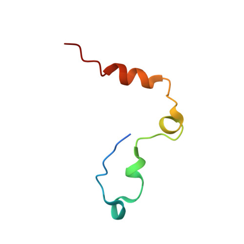
Prothrombin
A, C, E
49Homo sapiensMutation(s): 0 
Gene Names: F2
EC: 3.4.21.5
UniProt & NIH Common Fund Data Resources
Find proteins for P00734 (Homo sapiens)
Explore P00734 
Go to UniProtKB:  P00734
PHAROS:  P00734
GTEx:  ENSG00000180210 
Entity Groups  
Sequence Clusters30% Identity50% Identity70% Identity90% Identity95% Identity100% Identity
UniProt GroupP00734
Sequence Annotations
Expand
  • Reference Sequence
Find similar proteins by:  (by identity cutoff)  |  3D Structure
Entity ID: 2
MoleculeChains Sequence LengthOrganismDetailsImage
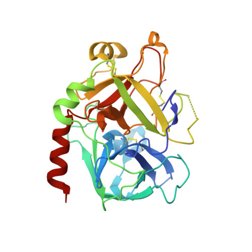
Prothrombin
B, D, F
259Homo sapiensMutation(s): 1 
Gene Names: F2
EC: 3.4.21.5
UniProt & NIH Common Fund Data Resources
Find proteins for P00734 (Homo sapiens)
Explore P00734 
Go to UniProtKB:  P00734
PHAROS:  P00734
GTEx:  ENSG00000180210 
Entity Groups  
Sequence Clusters30% Identity50% Identity70% Identity90% Identity95% Identity100% Identity
UniProt GroupP00734
Sequence Annotations
Expand
  • Reference Sequence
Find similar proteins by:  (by identity cutoff)  |  3D Structure
Entity ID: 3
MoleculeChains Sequence LengthOrganismDetailsImage
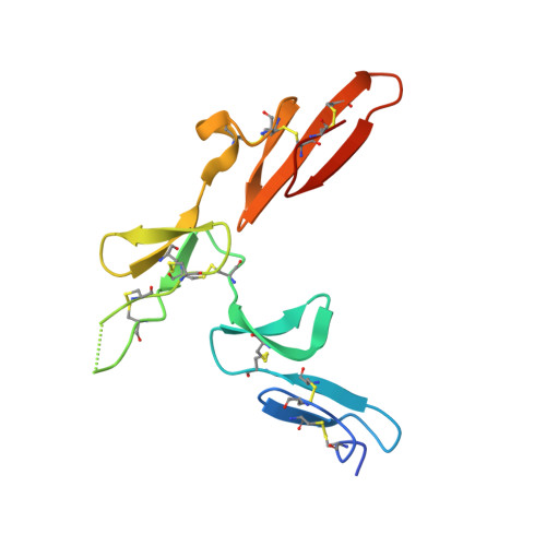
ThrombomodulinG [auth X],
H [auth Y],
I [auth Z]
121Homo sapiensMutation(s): 3 
Gene Names: THBDTHRM
UniProt & NIH Common Fund Data Resources
Find proteins for P07204 (Homo sapiens)
Explore P07204 
Go to UniProtKB:  P07204
PHAROS:  P07204
GTEx:  ENSG00000178726 
Entity Groups  
Sequence Clusters30% Identity50% Identity70% Identity90% Identity95% Identity100% Identity
UniProt GroupP07204
Sequence Annotations
Expand
  • Reference Sequence
Small Molecules
Ligands 2 Unique
IDChains Name / Formula / InChI Key2D Diagram3D Interactions
SO4
Query on SO4

Download Ideal Coordinates CCD File 
J [auth A]
K [auth A]
L [auth B]
M [auth B]
N [auth B]
J [auth A],
K [auth A],
L [auth B],
M [auth B],
N [auth B],
O [auth B],
P [auth B],
Q [auth C],
R [auth D],
S [auth D],
T [auth D],
U [auth D],
V [auth E],
W [auth F],
X [auth F]
SULFATE ION
O4 S
QAOWNCQODCNURD-UHFFFAOYSA-L
CA
Query on CA

Download Ideal Coordinates CCD File 
AA [auth Z],
Y [auth X],
Z [auth Y]
CALCIUM ION
Ca
BHPQYMZQTOCNFJ-UHFFFAOYSA-N
Experimental Data & Validation

Experimental Data

  • Method: X-RAY DIFFRACTION
  • Resolution: 2.40 Å
  • R-Value Free: 0.259 
  • R-Value Work: 0.211 
  • R-Value Observed: 0.243 
  • Space Group: P 21 21 21
Unit Cell:
Length ( Å )Angle ( ˚ )
a = 66.25α = 90
b = 100.34β = 90
c = 229.28γ = 90
Software Package:
Software NamePurpose
MOSFLMdata reduction
SCALAdata scaling
PHASERphasing
CNSrefinement
PDB_EXTRACTdata extraction
ADSCdata collection

Structure Validation

View Full Validation Report


Entry History 

Deposition Data

Revision History  (Full details and data files)

  • Version 1.0: 2009-08-18
    Type: Initial release
  • Version 1.1: 2011-07-13
    Changes: Version format compliance
  • Version 1.2: 2016-02-17
    Changes: Other
  • Version 1.3: 2023-11-01
    Changes: Data collection, Database references, Derived calculations, Refinement description