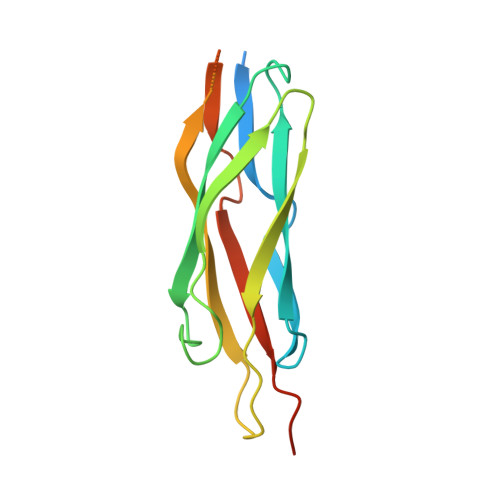
Structure and functional analysis of a Ca2+ sensor mutant of the na+/ca2+ exchanger
Chaptal, V., Ottolia, M., Mercado-Besserer, G., Nicoll, D.A., Philipson, K.D., Abramson, J.(2009) J Biological Chem 284: 14688-14692
- PubMed: 19332552 Search on PubMedSearch on PubMed Central
- DOI: https://doi.org/10.1074/jbc.C900037200
- Primary Citation Related Structures:
3GIN - PubMed Abstract:
The mammalian Na(+)/Ca(2+) exchanger, NCX1.1, serves as the main mechanism for Ca(2+) efflux across the sarcolemma following cardiac contraction. In addition to transporting Ca(2+), NCX1.1 activity is also strongly regulated by Ca(2+) binding to two intracellular regulatory domains, CBD1 and CBD2. The structures of both of these domains have been solved by NMR spectroscopy and x-ray crystallography, greatly enhancing our understanding of Ca(2+) regulation. Nevertheless, the mechanisms by which Ca(2+) regulates the exchanger remain incompletely understood. The initial NMR study showed that the first regulatory domain, CBD1, unfolds in the absence of regulatory Ca(2+). It was further demonstrated that a mutation of an acidic residue involved in Ca(2+) binding, E454K, prevents this structural unfolding. A contradictory result was recently obtained in a second NMR study in which Ca(2+) removal merely triggered local rearrangements of CBD1. To address this issue, we solved the crystal structure of the E454K-CBD1 mutant and performed electrophysiological analyses of the full-length exchanger with mutations at position 454. We show that the lysine substitution replaces the Ca(2+) ion at position 1 of the CBD1 Ca(2+) binding site and participates in a charge compensation mechanism. Electrophysiological analyses show that mutations of residue Glu-454 have no impact on Ca(2+) regulation of NCX1.1. Together, structural and mutational analyses indicate that only two of the four Ca(2+) ions that bind to CBD1 are important for regulating exchanger activity.
- Department of Physiology and Cardiovascular Research Laboratories, David Geffen School of Medicine at UCLA, Los Angeles, California 90095, USA.
Organizational Affiliation: