3GGP | pdb_00003ggp

Crystal structure of prephenate dehydrogenase from A. aeolicus in complex with hydroxyphenyl propionate and NAD+


Experimental Data Snapshot

  • Method: X-RAY DIFFRACTION
  • Resolution: 2.25 Å
  • R-Value Free: 
    0.248 (Depositor), 0.237 (DCC) 
  • R-Value Work: 
    0.198 (Depositor), 0.189 (DCC) 
  • R-Value Observed: 
    0.199 (Depositor) 

Starting Model: experimental
View more details

wwPDB Validation 3D Report Full Report

Validation slider image for 3GGP

Ligand Structure Quality Assessment 


This is version 1.2 of the entry. See complete history

Literature

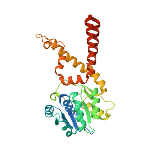
The Crystal Structure of Aquifex aeolicus Prephenate Dehydrogenase Reveals the Mode of Tyrosine Inhibition.

Sun, W.Shahinas, D.Bonvin, J.Hou, W.Kimber, M.S.Turnbull, J.Christendat, D.

(2009) J Biological Chem 284: 13223-13232

  • DOI: https://doi.org/10.1074/jbc.M806272200
  • Primary Citation Related Structures: 
    3GGG, 3GGO, 3GGP

  • PubMed Abstract: 

    TyrA proteins belong to a family of dehydrogenases that are dedicated to l-tyrosine biosynthesis. The three TyrA subclasses are distinguished by their substrate specificities, namely the prephenate dehydrogenases, the arogenate dehydrogenases, and the cyclohexadienyl dehydrogenases, which utilize prephenate, l-arogenate, or both substrates, respectively. The molecular mechanism responsible for TyrA substrate selectivity and regulation is unknown. To further our understanding of TyrA-catalyzed reactions, we have determined the crystal structures of Aquifex aeolicus prephenate dehydrogenase bound with NAD(+) plus either 4-hydroxyphenylpyuvate, 4-hydroxyphenylpropionate, or l-tyrosine and have used these structures as guides to target active site residues for site-directed mutagenesis. From a combination of mutational and structural analyses, we have demonstrated that His-147 and Arg-250 are key catalytic and binding groups, respectively, and Ser-126 participates in both catalysis and substrate binding through the ligand 4-hydroxyl group. The crystal structure revealed that tyrosine, a known inhibitor, binds directly to the active site of the enzyme and not to an allosteric site. The most interesting finding though, is that mutating His-217 relieved the inhibitory effect of tyrosine on A. aeolicus prephenate dehydrogenase. The identification of a tyrosine-insensitive mutant provides a novel avenue for designing an unregulated enzyme for application in metabolic engineering.


  • Organizational Affiliation
    • Department of Cell and Systems Biology, University of Toronto, Ontario, Canada.

Macromolecule Content 

  • Total Structure Weight: 144.11 kDa 
  • Atom Count: 9,369 
  • Modeled Residue Count: 1,142 
  • Deposited Residue Count: 1,256 
  • Unique protein chains: 1

Macromolecules

Find similar proteins by:|  3D Structure
Entity ID: 1
MoleculeChains  Sequence LengthOrganismDetailsImage
Prephenate dehydrogenase
A, B, C, D
314Aquifex aeolicusMutation(s): 0 
Gene Names: aq_1755tyrA
EC: 1.3.1.12
UniProt
Find proteins for O67636 (Aquifex aeolicus (strain VF5))
Explore O67636 
Go to UniProtKB:  O67636
Entity Groups
Sequence Clusters30% Identity50% Identity70% Identity90% Identity95% Identity100% Identity
UniProt GroupO67636
Sequence Annotations
Expand
Reference Sequence

Small Molecules

Ligands 3 Unique
IDChains Name / Formula / InChI Key2D Diagram3D Interactions
NAD

Query on NAD


Download:Ideal Coordinates CCD File
E [auth A],
G [auth B],
J [auth C],
L [auth D]
NICOTINAMIDE-ADENINE-DINUCLEOTIDE
C21 H27 N7 O14 P2
BAWFJGJZGIEFAR-NNYOXOHSSA-N
HPP

Query on HPP


Download:Ideal Coordinates CCD File
H [auth B],
K [auth C]
HYDROXYPHENYL PROPIONIC ACID
C9 H10 O3
NMHMNPHRMNGLLB-UHFFFAOYSA-N
CL

Query on CL


Download:Ideal Coordinates CCD File
F [auth A],
I [auth B]
CHLORIDE ION
Cl
VEXZGXHMUGYJMC-UHFFFAOYSA-M

Experimental Data & Validation

Experimental Data

  • Method: X-RAY DIFFRACTION
  • Resolution: 2.25 Å
  • R-Value Free:  0.248 (Depositor), 0.237 (DCC) 
  • R-Value Work:  0.198 (Depositor), 0.189 (DCC) 
  • R-Value Observed: 0.199 (Depositor) 
Space Group: P 21 21 21
Unit Cell:
Length ( Å )Angle ( ˚ )
a = 82.061α = 90
b = 92.361β = 90
c = 163.83γ = 90
Software Package:
Software NamePurpose
HKL-3000data collection
AMoREphasing
REFMACrefinement
HKL-3000data reduction
HKL-3000data scaling

Structure Validation

View Full Validation Report


Ligand Structure Quality Assessment 


Entry History 

Deposition Data

Revision History  (Full details and data files)

  • Version 1.0: 2009-03-10
    Type: Initial release
  • Version 1.1: 2011-07-13
    Changes: Advisory, Refinement description, Version format compliance
  • Version 1.2: 2023-09-06
    Changes: Data collection, Database references, Derived calculations, Refinement description