3GDD | pdb_00003gdd

An inverted anthraquinone-DNA crystal structure


Experimental Data Snapshot

  • Method: X-RAY DIFFRACTION
  • Resolution: 2.80 Å
  • R-Value Free: 
    0.259 (Depositor), 0.269 (DCC) 
  • R-Value Work: 
    0.232 (Depositor), 0.252 (DCC) 
  • R-Value Observed: 
    0.233 (Depositor) 

wwPDB Validation 3D Report Full Report

Validation slider image for 3GDD

Ligand Structure Quality Assessment 


This is version 1.2 of the entry. See complete history

Literature

Structure of a stacked anthraquinone-DNA complex

De Luchi, D.Uson, I.Wright, G.Gouyette, C.Subirana, J.A.

(2010) Acta Crystallogr Sect F Struct Biol Cryst Commun 66: 1019-1022

  • DOI: https://doi.org/10.1107/S1744309110030034
  • Primary Citation Related Structures: 
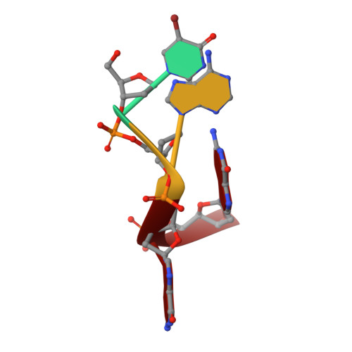
    3GDD

  • PubMed Abstract: 

    The crystal structure of the telomeric sequence d(UBrAGG) interacting with an anthraquinone derivative has been solved by MAD. In all previously studied complexes of intercalating drugs, the drug is usually sandwiched between two DNA base pairs. Instead, the present structure looks like a crystal of stacked anthraquinone molecules in which isolated base pairs are intercalated. Unusual base pairs are present in the structure, such as G.G and A.UBr reverse Watson-Crick base pairs.


  • Organizational Affiliation
    • Departament d'Enginyeria Química, Universitat Politècnica de Catalunya, Spain.

Macromolecule Content 

  • Total Structure Weight: 4.25 kDa 
  • Atom Count: 259 
  • Modeled Residue Count: 8 
  • Deposited Residue Count: 8 
  • Unique nucleic acid chains: 1

Macromolecules

Find similar nucleic acids by:  Sequence
Entity ID: 1
MoleculeChains LengthOrganismImage
5'-D(*(BRU)P*AP*GP*G)-3'
A, B
4N/A
Sequence Annotations
Expand
Reference Sequence

Experimental Data & Validation

Experimental Data

  • Method: X-RAY DIFFRACTION
  • Resolution: 2.80 Å
  • R-Value Free:  0.259 (Depositor), 0.269 (DCC) 
  • R-Value Work:  0.232 (Depositor), 0.252 (DCC) 
  • R-Value Observed: 0.233 (Depositor) 
Space Group: P 43 21 2
Unit Cell:
Length ( Å )Angle ( ˚ )
a = 29.698α = 90
b = 29.698β = 90
c = 60.883γ = 90
Software Package:
Software NamePurpose
SOLVEphasing
REFMACrefinement
HKL-2000data reduction
HKL-2000data scaling

Structure Validation

View Full Validation Report


Ligand Structure Quality Assessment 


Entry History 

Deposition Data

Revision History  (Full details and data files)

  • Version 1.0: 2010-03-09
    Type: Initial release
  • Version 1.1: 2011-07-13
    Changes: Version format compliance
  • Version 1.2: 2024-02-21
    Changes: Data collection, Database references, Derived calculations