3GCZ | pdb_00003gcz

Yokose virus Methyltransferase in complex with AdoMet


Experimental Data Snapshot

  • Method: X-RAY DIFFRACTION
  • Resolution: 1.70 Å
  • R-Value Free: 
    0.198 (Depositor), 0.193 (DCC) 
  • R-Value Work: 
    0.167 (Depositor), 0.172 (DCC) 
  • R-Value Observed: 
    0.169 (Depositor) 

Starting Model: experimental
View more details

wwPDB Validation 3D Report Full Report

Validation slider image for 3GCZ

Ligand Structure Quality Assessment 


This is version 1.2 of the entry. See complete history

Literature

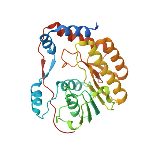
Crystal structure of a methyltransferase from a no-known-vector Flavivirus

Bollati, M.Milani, M.Mastrangelo, E.de Lambellarie, X.Canard, B.Bolognesi, M.

(2009) Biochem Biophys Res Commun 382: 200-204

  • DOI: https://doi.org/10.1016/j.bbrc.2009.03.008
  • Primary Citation Related Structures: 
    3GCZ

  • PubMed Abstract: 

    Presently known flaviviruses belong to three major evolutionary branches: tick-borne viruses, mosquito-borne viruses and viruses with no known vector. Here we present the crystal structure of the Yokose virus methyltransferase at 1.7A resolution, the first structure of a methyltransferase of a Flavivirus with no known vector. Structural comparison of three methyltransferases representative of each of the Flavivirus branches shows that fold and structures are closely conserved, most differences being related to surface loops flexibility. Analysis of the conserved residues throughout all the sequenced flaviviral methyltransferases reveals that, besides the central cleft hosting the substrate and cofactor binding sites, a second, almost continuous, patch is conserved and points away from active site towards the back of the protein. The high level of structural conservation in this region could be functional for the methyltransferase/RNA interaction and stabilization of the ensuing complex.


  • Organizational Affiliation
    • Department of Biomolecular Sciences and Biotechnology, CNR-INFM and CIMAINA, University of Milano, Via Celoria 26, 20133 Milano, Italy.

Macromolecule Content 

  • Total Structure Weight: 32.61 kDa 
  • Atom Count: 2,272 
  • Modeled Residue Count: 259 
  • Deposited Residue Count: 282 
  • Unique protein chains: 1

Macromolecules

Find similar proteins by:|  3D Structure
Entity ID: 1
MoleculeChains  Sequence LengthOrganismDetailsImage
Polyprotein282Yokose virusMutation(s): 0 
EC: 2.1.1.57
UniProt
Find proteins for Q7T918 (Yokose virus)
Explore Q7T918 
Go to UniProtKB:  Q7T918
Entity Groups
Sequence Clusters30% Identity50% Identity70% Identity90% Identity95% Identity100% Identity
UniProt GroupQ7T918
Sequence Annotations
Expand
Reference Sequence

Small Molecules

Ligands 3 Unique
IDChains Name / Formula / InChI Key2D Diagram3D Interactions
SAM

Query on SAM


Download:Ideal Coordinates CCD File
B [auth A]S-ADENOSYLMETHIONINE
C15 H22 N6 O5 S
MEFKEPWMEQBLKI-FCKMPRQPSA-N
SO4

Query on SO4


Download:Ideal Coordinates CCD File
F [auth A],
G [auth A],
H [auth A]
SULFATE ION
O4 S
QAOWNCQODCNURD-UHFFFAOYSA-L
GOL

Query on GOL


Download:Ideal Coordinates CCD File
C [auth A],
D [auth A],
E [auth A]
GLYCEROL
C3 H8 O3
PEDCQBHIVMGVHV-UHFFFAOYSA-N

Experimental Data & Validation

Experimental Data

  • Method: X-RAY DIFFRACTION
  • Resolution: 1.70 Å
  • R-Value Free:  0.198 (Depositor), 0.193 (DCC) 
  • R-Value Work:  0.167 (Depositor), 0.172 (DCC) 
  • R-Value Observed: 0.169 (Depositor) 
Space Group: P 32
Unit Cell:
Length ( Å )Angle ( ˚ )
a = 68.912α = 90
b = 68.912β = 90
c = 50.806γ = 120
Software Package:
Software NamePurpose
MOSFLMdata reduction
MOLREPphasing
REFMACrefinement
SCALAdata scaling

Structure Validation

View Full Validation Report


Ligand Structure Quality Assessment 


Entry History 

Deposition Data

Revision History  (Full details and data files)

  • Version 1.0: 2009-03-24
    Type: Initial release
  • Version 1.1: 2011-07-13
    Changes: Advisory, Version format compliance
  • Version 1.2: 2023-11-01
    Changes: Data collection, Database references, Derived calculations, Refinement description