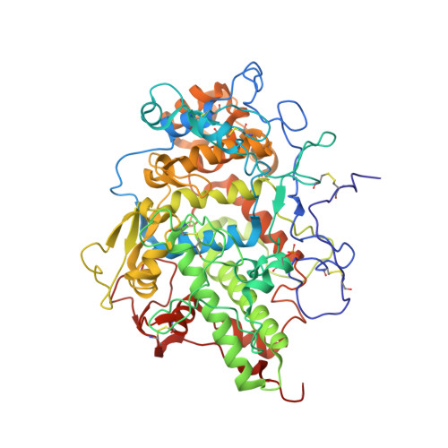
Mode of ligand binding and assignment of subsites in mammalian peroxidases: crystal structure of lactoperoxidase complexes with acetyl salycylic acid, salicylhydroxamic acid and benzylhydroxamic acid
Singh, A.K., Singh, N., Sinha, M., Bhushan, A., Kaur, P., Srinivasan, A., Sharma, S., Singh, T.P.To be published.