3G80 | pdb_00003g80

Nodamura virus protein b2, RNA-binding domain


Experimental Data Snapshot

  • Method: X-RAY DIFFRACTION
  • Resolution: 2.50 Å
  • R-Value Free: 
    0.297 (Depositor), 0.286 (DCC) 
  • R-Value Work: 
    0.233 (Depositor), 0.226 (DCC) 
  • R-Value Observed: 
    0.233 (Depositor) 

Starting Model: experimental
View more details

wwPDB Validation 3D Report Full Report

Validation slider image for 3G80

This is version 1.2 of the entry. See complete history

Literature

Structure of the RNA-Binding Domain of Nodamura Virus Protein B2, a Suppressor of RNA Interference.

Korber, S.Shaik Syed Ali, P.Chen, J.C.

(2009) Biochemistry 48: 2307-2309

  • DOI: https://doi.org/10.1021/bi900126s
  • Primary Citation Related Structures: 
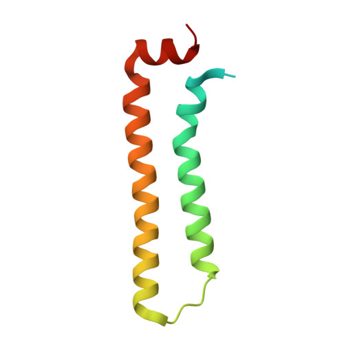
    3G80

  • PubMed Abstract: 

    Protein B2 from Nodamura virus (NMV B2), a member of the Nodavirus family, acts as a suppressor of RNA interference (RNAi). The N-terminal domain of NMV B2, consisting of residues 1-79, recognizes double-stranded RNA (dsRNA). The 2.5 A crystal structure of the RNA-binding domain of NMV B2 shows a dimeric, helical bundle structure. The structure shows a conserved set of RNA-binding residues compared with flock house virus B2, despite limited sequence identity. The crystal packing places the RNA-binding residues along one face of symmetry-related molecules, suggesting a potential platform for recognition of dsRNA.


  • Organizational Affiliation
    • Institute of Biophysical Chemistry, Goethe University Frankfurt, Max-von-Laue-Strasse 9, 60438 Frankfurt, Germany.

Macromolecule Content 

  • Total Structure Weight: 22.27 kDa 
  • Atom Count: 1,223 
  • Modeled Residue Count: 146 
  • Deposited Residue Count: 194 
  • Unique protein chains: 1

Macromolecules

Find similar proteins by:|  3D Structure
Entity ID: 1
MoleculeChains  Sequence LengthOrganismDetailsImage
Protein B2
A, B
97Nodamura virusMutation(s): 0 
Gene Names: B2
UniProt
Find proteins for Q9IMM3 (Nodamura virus (strain Mag115))
Explore Q9IMM3 
Go to UniProtKB:  Q9IMM3
Entity Groups
Sequence Clusters30% Identity50% Identity70% Identity90% Identity95% Identity100% Identity
UniProt GroupQ9IMM3
Sequence Annotations
Expand
Reference Sequence

Experimental Data & Validation

Experimental Data

  • Method: X-RAY DIFFRACTION
  • Resolution: 2.50 Å
  • R-Value Free:  0.297 (Depositor), 0.286 (DCC) 
  • R-Value Work:  0.233 (Depositor), 0.226 (DCC) 
  • R-Value Observed: 0.233 (Depositor) 
Space Group: P 21 21 21
Unit Cell:
Length ( Å )Angle ( ˚ )
a = 32.19α = 90
b = 56.6β = 90
c = 98.56γ = 90
Software Package:
Software NamePurpose
MOLREPphasing
CNSrefinement
PDB_EXTRACTdata extraction
ADSCdata collection
MOSFLMdata reduction
SCALAdata scaling

Structure Validation

View Full Validation Report


Entry History 

Deposition Data

Revision History  (Full details and data files)

  • Version 1.0: 2010-01-05
    Type: Initial release
  • Version 1.1: 2011-07-13
    Changes: Version format compliance
  • Version 1.2: 2023-09-06
    Changes: Data collection, Database references, Refinement description