3G6H | pdb_00003g6h

Src Thr338Ile inhibited in the DFG-Asp-Out conformation


Experimental Data Snapshot

  • Method: X-RAY DIFFRACTION
  • Resolution: 2.35 Å
  • R-Value Free: 
    0.283 (Depositor), 0.278 (DCC) 
  • R-Value Work: 
    0.225 (Depositor), 0.220 (DCC) 
  • R-Value Observed: 
    0.228 (Depositor) 

Starting Model: experimental
View more details

wwPDB Validation 3D Report Full Report

Validation slider image for 3G6H

Ligand Structure Quality Assessment 


This is version 1.3 of the entry. See complete history

Literature

Equally potent inhibition of c-Src and Abl by compounds that recognize inactive kinase conformations

Seeliger, M.A.Ranjitkar, P.Kasap, C.Shan, Y.Shaw, D.E.Shah, N.P.Kuriyan, J.Maly, D.J.

(2009) Cancer Res 69: 2384-2392

  • DOI: https://doi.org/10.1158/0008-5472.CAN-08-3953
  • Primary Citation Related Structures: 
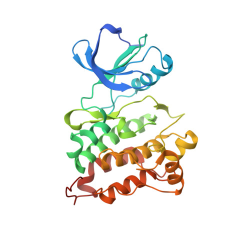
    3G6G, 3G6H

  • PubMed Abstract: 

    Imatinib is an inhibitor of the Abl tyrosine kinase domain that is effective in the treatment of chronic myelogenic leukemia. Although imatinib binds tightly to the Abl kinase domain, its affinity for the closely related kinase domain of c-Src is at least 2,000-fold lower. Imatinib recognition requires a specific inactive conformation of the kinase domain, in which a conserved Asp-Phe-Gly (DFG) motif is flipped with respect to the active conformation. The inability of c-Src to readily adopt this flipped DFG conformation was thought to underlie the selectivity of imatinib for Abl over c-Src. Here, we present a series of inhibitors (DSA compounds) that are based on the core scaffold of imatinib but which bind with equally high potency to c-Src and Abl. The DSA compounds bind to c-Src in the DFG-flipped conformation, as confirmed by crystal structures and kinetic analysis. The origin of the high affinity of these compounds for c-Src is suggested by the fact that they also inhibit clinically relevant Abl variants bearing mutations in a structural element, the P-loop, that normally interacts with the phosphate groups of ATP but is folded over a substructure of imatinib in Abl. Importantly, several of the DSA compounds block the growth of Ba/F3 cells harboring imatinib-resistant BCR-ABL mutants, including the Thr315Ile "gatekeeper" mutation, but do not suppress the growth of parental Ba/F3 cells.


  • Organizational Affiliation
    • Howard Hughes Medical Institute and Department of Molecular and Cell Biology, University of California, Berkely, USA.

Macromolecule Content 

  • Total Structure Weight: 66.11 kDa 
  • Atom Count: 4,384 
  • Modeled Residue Count: 525 
  • Deposited Residue Count: 572 
  • Unique protein chains: 1

Macromolecules

Find similar proteins by:|  3D Structure
Entity ID: 1
MoleculeChains  Sequence LengthOrganismDetailsImage
Proto-oncogene tyrosine-protein kinase Src
A, B
286Gallus gallusMutation(s): 1 
Gene Names: SRC
EC: 2.7.10.2
UniProt
Find proteins for P00523 (Gallus gallus)
Explore P00523 
Go to UniProtKB:  P00523
Entity Groups
Sequence Clusters30% Identity50% Identity70% Identity90% Identity95% Identity100% Identity
UniProt GroupP00523
Sequence Annotations
Expand
Reference Sequence

Small Molecules

Ligands 1 Unique
IDChains Name / Formula / InChI Key2D Diagram3D Interactions
G6H

Query on G6H


Download:Ideal Coordinates CCD File
C [auth B]N-{4-methyl-3-[(3-{4-[(3,4,5-trimethoxyphenyl)amino]-1,3,5-triazin-2-yl}pyridin-2-yl)amino]phenyl}-3-(trifluoromethyl)benzamide
C32 H28 F3 N7 O4
SNOVXGXKOHJZJH-UHFFFAOYSA-N

Experimental Data & Validation

Experimental Data

  • Method: X-RAY DIFFRACTION
  • Resolution: 2.35 Å
  • R-Value Free:  0.283 (Depositor), 0.278 (DCC) 
  • R-Value Work:  0.225 (Depositor), 0.220 (DCC) 
  • R-Value Observed: 0.228 (Depositor) 
Space Group: P 1
Unit Cell:
Length ( Å )Angle ( ˚ )
a = 41.938α = 100.99
b = 63.344β = 89.39
c = 74.179γ = 90.2
Software Package:
Software NamePurpose
HKL-2000data collection
PHASERphasing
REFMACrefinement
HKL-2000data reduction
SCALEPACKdata scaling

Structure Validation

View Full Validation Report


Ligand Structure Quality Assessment 


Entry History 

Deposition Data

Revision History  (Full details and data files)

  • Version 1.0: 2009-03-24
    Type: Initial release
  • Version 1.1: 2011-07-13
    Changes: Version format compliance
  • Version 1.2: 2021-10-20
    Changes: Database references, Derived calculations
  • Version 1.3: 2023-09-06
    Changes: Data collection, Refinement description