3FZW

Crystal Structure of Ketosteroid Isomerase D40N-D103N from Pseudomonas putida (pKSI) with bound equilenin


Experimental Data Snapshot

  • Method: X-RAY DIFFRACTION
  • Resolution: 1.32 Å
  • R-Value Free: 0.183 
  • R-Value Work: 0.149 
  • R-Value Observed: 0.151 

wwPDB Validation   3D Report Full Report


Ligand Structure Quality Assessment 


This is version 1.4 of the entry. See complete history


Literature

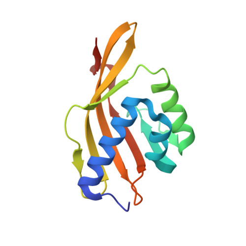
Hydrogen bond coupling in the ketosteroid isomerase active site.

Sigala, P.A.Caaveiro, J.M.Ringe, D.Petsko, G.A.Herschlag, D.

(2009) Biochemistry 48: 6932-6939

  • DOI: https://doi.org/10.1021/bi900713j
  • Primary Citation of Related Structures:  
    3FZW

  • PubMed Abstract: 

    Hydrogen bond networks are key elements of biological structure and function. Nevertheless, their structural properties are challenging to assess within complex macromolecules. Hydrogen-bonded protons are not observed in the vast majority of protein X-ray structures, and static crystallographic models provide limited information regarding the dynamical coupling within hydrogen bond networks. We have brought together 1.1-1.3 A resolution X-ray crystallography, (1)H NMR, site-directed mutagenesis, and deuterium isotope effects on the geometry and chemical shifts of hydrogen-bonded protons to probe the conformational coupling of hydrogen bonds donated by Y16 and D103 in the oxyanion hole of bacterial ketosteroid isomerase. Our results suggest a robust physical coupling of the equilibrium structures of these two hydrogen bonds such that a lengthening of one hydrogen bond by as little as 0.01 A results in a shortening of the neighbor by a similar magnitude. Furthermore, the structural rearrangements detected by NMR in response to mutations within the active site hydrogen bond network can be explained on the basis of the observed coupling. The results herein elucidate fundamental structural properties of hydrogen bonds within the idiosyncratic environment of an enzyme active site and provide a foundation for future experimental and computational explorations of the role of coupled motions within hydrogen bond networks.


  • Organizational Affiliation

    Department of Biochemistry, Stanford University, Stanford, California 94305, USA.


Macromolecules
Find similar proteins by:  (by identity cutoff)  |  3D Structure
Entity ID: 1
MoleculeChains Sequence LengthOrganismDetailsImage
Steroid Delta-isomerase
A, B
131Pseudomonas putidaMutation(s): 2 
Gene Names: ksi
EC: 5.3.3.1
UniProt
Find proteins for P07445 (Pseudomonas putida)
Explore P07445 
Go to UniProtKB:  P07445
Entity Groups  
Sequence Clusters30% Identity50% Identity70% Identity90% Identity95% Identity100% Identity
UniProt GroupP07445
Sequence Annotations
Expand
  • Reference Sequence
Experimental Data & Validation

Experimental Data

  • Method: X-RAY DIFFRACTION
  • Resolution: 1.32 Å
  • R-Value Free: 0.183 
  • R-Value Work: 0.149 
  • R-Value Observed: 0.151 
  • Space Group: C 1 2 1
Unit Cell:
Length ( Å )Angle ( ˚ )
a = 86.876α = 90
b = 71.196β = 89.92
c = 50.452γ = 90
Software Package:
Software NamePurpose
DENZOdata reduction
SCALEPACKdata scaling
MOLREPphasing
REFMACrefinement
PDB_EXTRACTdata extraction
ADSCdata collection

Structure Validation

View Full Validation Report


Ligand Structure Quality Assessment 


Entry History 

Deposition Data

Revision History  (Full details and data files)

  • Version 1.0: 2009-06-02
    Type: Initial release
  • Version 1.1: 2011-07-13
    Changes: Non-polymer description, Version format compliance
  • Version 1.2: 2017-11-01
    Changes: Refinement description
  • Version 1.3: 2021-10-20
    Changes: Database references, Derived calculations
  • Version 1.4: 2023-09-06
    Changes: Data collection, Refinement description