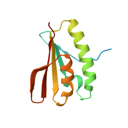
Experimental Data Snapshot

  • Method: X-RAY DIFFRACTION
  • Resolution: 2.30 Å
  • R-Value Free: 
    0.235 (Depositor), 0.262 (DCC) 
  • R-Value Work: 
    0.201 (Depositor), 0.227 (DCC) 
  • R-Value Observed: 
    0.203 (Depositor) 

wwPDB Validation 3D Report Full Report

Validation slider image for 3FXT

This is version 1.2 of the entry. See complete history

Macromolecule Content 

  • Total Structure Weight: 101.73 kDa 
  • Atom Count: 6,088 
  • Modeled Residue Count: 711 
  • Deposited Residue Count: 904 
  • Unique protein chains: 1

Macromolecules

Find similar proteins by:|  3D Structure
Entity ID: 1
MoleculeChains  Sequence LengthOrganismDetailsImage
Nucleoside diphosphate-linked moiety X motif 6
A, B, C, D, E
A, B, C, D, E, F, G, H
113Homo sapiensMutation(s): 0 
Gene Names: FGF2ASNUDT6
EC: 3.6.1.18 (UniProt), 3.6.1.22 (UniProt)
UniProt & NIH Common Fund Data Resources
Find proteins for P53370 (Homo sapiens)
Explore P53370 
Go to UniProtKB:  P53370
PHAROS:  P53370
GTEx:  ENSG00000170917 
Entity Groups
Sequence Clusters30% Identity50% Identity70% Identity90% Identity95% Identity100% Identity
UniProt GroupP53370
Sequence Annotations
Expand
Reference Sequence

Experimental Data & Validation

Experimental Data

  • Method: X-RAY DIFFRACTION
  • Resolution: 2.30 Å
  • R-Value Free:  0.235 (Depositor), 0.262 (DCC) 
  • R-Value Work:  0.201 (Depositor), 0.227 (DCC) 
  • R-Value Observed: 0.203 (Depositor) 
Space Group: P 64 2 2
Unit Cell:
Length ( Å )Angle ( ˚ )
a = 185.8α = 90
b = 185.8β = 90
c = 129.2γ = 120
Software Package:
Software NamePurpose
ADSCdata collection
autoSHARPphasing
REFMACrefinement
XDSdata reduction
XSCALEdata scaling

Structure Validation

View Full Validation Report


Entry History 

Revision History  (Full details and data files)

  • Version 1.0: 2009-03-03
    Type: Initial release
  • Version 1.1: 2011-07-13
    Changes: Advisory, Version format compliance
  • Version 1.2: 2024-03-20
    Changes: Data collection, Database references, Derived calculations