3FPD

G9a-like protein lysine methyltransferase inhibition by BIX-01294


Experimental Data Snapshot

  • Method: X-RAY DIFFRACTION
  • Resolution: 2.40 Å
  • R-Value Free: 0.262 
  • R-Value Work: 0.221 

wwPDB Validation   3D Report Full Report


Ligand Structure Quality Assessment 


This is version 1.2 of the entry. See complete history


Literature

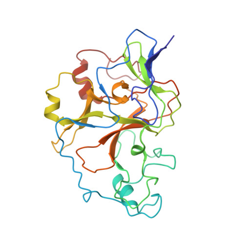
Structural basis for G9a-like protein lysine methyltransferase inhibition by BIX-01294.

Chang, Y.Zhang, X.Horton, J.R.Upadhyay, A.K.Spannhoff, A.Liu, J.Snyder, J.P.Bedford, M.T.Cheng, X.

(2009) Nat Struct Mol Biol 16: 312-317

  • DOI: https://doi.org/10.1038/nsmb.1560
  • Primary Citation of Related Structures:  
    3FPD

  • PubMed Abstract: 
  • Histone lysine methylation is an important epigenetic mark that regulates gene expression and chromatin organization. G9a and G9a-like protein (GLP) are euchromatin-associated methyltransferases that repress transcription by methylating histone H3 Lys9. BIX-01294 was originally identified as a G9a inhibitor during a chemical library screen of small molecules and has previously been used in the generation of induced pluripotent stem cells ...

    Histone lysine methylation is an important epigenetic mark that regulates gene expression and chromatin organization. G9a and G9a-like protein (GLP) are euchromatin-associated methyltransferases that repress transcription by methylating histone H3 Lys9. BIX-01294 was originally identified as a G9a inhibitor during a chemical library screen of small molecules and has previously been used in the generation of induced pluripotent stem cells. Here we present the crystal structure of the catalytic SET domain of GLP in complex with BIX-01294 and S-adenosyl-L-homocysteine. The inhibitor is bound in the substrate peptide groove at the location where the histone H3 residues N-terminal to the target lysine lie in the previously solved structure of the complex with histone peptide. The inhibitor resembles the bound conformation of histone H3 Lys4 to Arg8, and is positioned in place by residues specific for G9a and GLP through specific interactions.


    Organizational Affiliation

    Department of Biochemistry, Emory University School of Medicine, 1510 Clifton Road, Atlanta, Georgia 30322, USA.


Macromolecules
Find similar proteins by:  (by identity cutoff)  |  3D Structure
Entity ID: 1
MoleculeChainsSequence LengthOrganismDetailsImage
Histone-lysine N-methyltransferase, H3 lysine-9 specific 5
A, B
261Homo sapiensMutation(s): 0 
Gene Names: EHMT1EUHMTASE1KIAA1876KMT1DGLP
EC: 2.1.1.43 (PDB Primary Data), 2.1.1 (UniProt), 2.1.1.367 (UniProt)
UniProt & NIH Common Fund Data Resources
Find proteins for Q9H9B1 (Homo sapiens)
Explore Q9H9B1 
Go to UniProtKB:  Q9H9B1
PHAROS:  Q9H9B1
Entity Groups  
Sequence Clusters30% Identity50% Identity70% Identity90% Identity95% Identity100% Identity
UniProt GroupQ9H9B1
Protein Feature View
Expand
  • Reference Sequence
Small Molecules
Binding Affinity Annotations 
IDSourceBinding Affinity
Q4A Binding MOAD:  3FPD IC50: 700 (nM) from 1 assay(s)
BindingDB:  3FPD IC50: min: 0.7, max: 700 (nM) from 4 assay(s)
PDBBind:  3FPD IC50: 700 (nM) from 1 assay(s)
SAH BindingDB:  3FPD IC50: 230 (nM) from 1 assay(s)
Experimental Data & Validation

Experimental Data

  • Method: X-RAY DIFFRACTION
  • Resolution: 2.40 Å
  • R-Value Free: 0.262 
  • R-Value Work: 0.221 
  • Space Group: P 21 21 21
Unit Cell:
Length ( Å )Angle ( ˚ )
a = 75.3α = 90
b = 95.2β = 90
c = 101.7γ = 90
Software Package:
Software NamePurpose
HKL-2000data collection
PHASERphasing
CNSrefinement
HKL-2000data reduction
HKL-2000data scaling

Structure Validation

View Full Validation Report


Ligand Structure Quality Assessment 


Entry History 

Deposition Data

Revision History  (Full details and data files)

  • Version 1.0: 2009-02-17
    Type: Initial release
  • Version 1.1: 2011-07-13
    Changes: Version format compliance
  • Version 1.2: 2023-09-06
    Changes: Data collection, Database references, Derived calculations, Refinement description