3FLO | pdb_00003flo

Crystal structure of the carboxyl-terminal domain of yeast DNA polymerase alpha in complex with its B subunit


Experimental Data Snapshot

  • Method: X-RAY DIFFRACTION
  • Resolution: 2.50 Å
  • R-Value Free: 
    0.218 (Depositor), 0.240 (DCC) 
  • R-Value Work: 
    0.191 (Depositor), 0.210 (DCC) 
  • R-Value Observed: 
    0.192 (Depositor) 

wwPDB Validation   3D Report Full Report


This is version 1.3 of the entry. See complete history


Literature

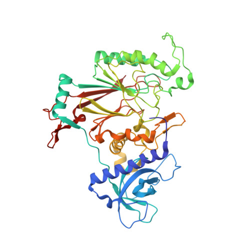
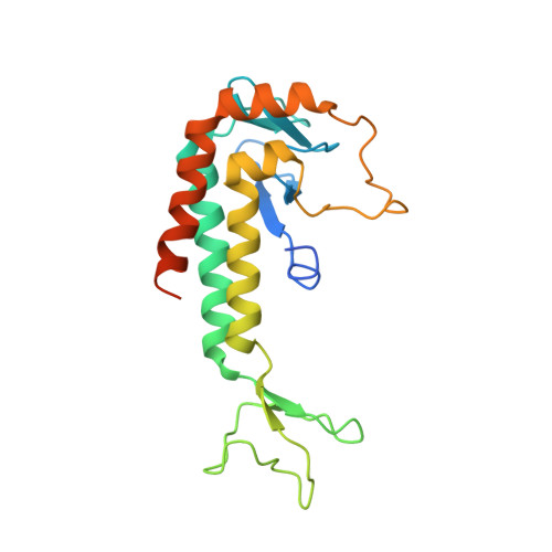
3D architecture of DNA Pol alpha reveals the functional core of multi-subunit replicative polymerases

Klinge, S.Nunez-Ramirez, R.Llorca, O.Pellegrini, L.

(2009) EMBO J 28: 1978-1987

  • DOI: https://doi.org/10.1038/emboj.2009.150
  • Primary Citation of Related Structures:  
    3FLO

  • PubMed Abstract: 

    Eukaryotic DNA replication requires the coordinated activity of the multi-subunit DNA polymerases: Pol alpha, Pol delta and Pol epsilon. The conserved catalytic and regulatory B subunits associate in a constitutive heterodimer that represents the functional core of all three replicative polymerases. Here, we combine X-ray crystallography and electron microscopy (EM) to describe subunit interaction and 3D architecture of heterodimeric yeast Pol alpha. The crystal structure of the C-terminal domain (CTD) of the catalytic subunit bound to the B subunit illustrates a conserved mechanism of accessory factor recruitment by replicative polymerases. The EM reconstructions of Pol alpha reveal a bilobal shape with separate catalytic and regulatory modules. Docking of the B-CTD complex in the EM reconstruction shows that the B subunit is tethered to the polymerase domain through a structured but flexible linker. Our combined findings provide a structural template for the common functional architecture of the three major replicative DNA polymerases.


  • Organizational Affiliation

    Department of Biochemistry, University of Cambridge, Cambridge, UK.


Macromolecules
Find similar proteins by:  (by identity cutoff)  |  3D Structure
Entity ID: 1
MoleculeChains Sequence LengthOrganismDetailsImage
DNA polymerase alpha subunit BA,
D [auth C],
G [auth E],
J [auth G]
460Saccharomyces cerevisiaeMutation(s): 0 
Gene Names: POL12
UniProt
Find proteins for P38121 (Saccharomyces cerevisiae (strain ATCC 204508 / S288c))
Explore P38121 
Go to UniProtKB:  P38121
Entity Groups  
Sequence Clusters30% Identity50% Identity70% Identity90% Identity95% Identity100% Identity
UniProt GroupP38121
Sequence Annotations
Expand
  • Reference Sequence

Find similar proteins by:  Sequence   |   3D Structure  

Entity ID: 2
MoleculeChains Sequence LengthOrganismDetailsImage
DNA polymerase alpha catalytic subunit AB [auth I],
E [auth J],
H [auth K],
K [auth L]
3Saccharomyces cerevisiaeMutation(s): 0 
Gene Names: POL1CDC17
Sequence Annotations
Expand
  • Reference Sequence
Find similar proteins by:  (by identity cutoff)  |  3D Structure
Entity ID: 3
MoleculeChains Sequence LengthOrganismDetailsImage
DNA polymerase alpha catalytic subunit AC [auth B],
F [auth D],
I [auth F],
L [auth H]
206Saccharomyces cerevisiaeMutation(s): 0 
Gene Names: POL1CDC17
EC: 2.7.7.7
UniProt
Find proteins for P13382 (Saccharomyces cerevisiae (strain ATCC 204508 / S288c))
Explore P13382 
Go to UniProtKB:  P13382
Entity Groups  
Sequence Clusters30% Identity50% Identity70% Identity90% Identity95% Identity100% Identity
UniProt GroupP13382
Sequence Annotations
Expand
  • Reference Sequence
Small Molecules
Ligands 2 Unique
IDChains Name / Formula / InChI Key2D Diagram3D Interactions
SO4
Query on SO4

Download Ideal Coordinates CCD File 
AB [auth E]
AC [auth G]
BA [auth B]
BB [auth E]
CA [auth B]
AB [auth E],
AC [auth G],
BA [auth B],
BB [auth E],
CA [auth B],
CB [auth E],
DA [auth B],
DB [auth E],
DC [auth H],
EA [auth B],
EB [auth E],
EC [auth H],
FA [auth B],
FB [auth E],
FC [auth H],
GA [auth B],
GB [auth E],
GC [auth H],
HA [auth C],
HB [auth E],
IA [auth C],
IB [auth E],
JA [auth C],
JB [auth E],
KA [auth C],
LA [auth C],
M [auth A],
MA [auth C],
MB [auth F],
N [auth A],
NA [auth C],
NB [auth F],
O [auth A],
OA [auth C],
OB [auth F],
P [auth A],
PA [auth C],
PB [auth F],
Q [auth A],
QA [auth C],
QB [auth G],
R [auth A],
RA [auth C],
RB [auth G],
S [auth A],
SB [auth G],
T [auth A],
TB [auth G],
U [auth A],
UA [auth D],
UB [auth G],
V [auth A],
VA [auth D],
VB [auth G],
W [auth A],
WA [auth D],
WB [auth G],
X [auth A],
XA [auth D],
XB [auth G],
Y [auth A],
YA [auth E],
YB [auth G],
ZA [auth E],
ZB [auth G]
SULFATE ION
O4 S
QAOWNCQODCNURD-UHFFFAOYSA-L
ZN
Query on ZN

Download Ideal Coordinates CCD File 
AA [auth B]
BC [auth H]
CC [auth H]
KB [auth F]
LB [auth F]
AA [auth B],
BC [auth H],
CC [auth H],
KB [auth F],
LB [auth F],
SA [auth D],
TA [auth D],
Z [auth B]
ZINC ION
Zn
PTFCDOFLOPIGGS-UHFFFAOYSA-N
Experimental Data & Validation

Experimental Data

  • Method: X-RAY DIFFRACTION
  • Resolution: 2.50 Å
  • R-Value Free:  0.218 (Depositor), 0.240 (DCC) 
  • R-Value Work:  0.191 (Depositor), 0.210 (DCC) 
  • R-Value Observed: 0.192 (Depositor) 
Space Group: P 1 21 1
Unit Cell:
Length ( Å )Angle ( ˚ )
a = 85.411α = 90
b = 142.633β = 102.33
c = 175.255γ = 90
Software Package:
Software NamePurpose
ADSCdata collection
PHENIXmodel building
REFMACrefinement
MOSFLMdata reduction
SCALAdata scaling
PHENIXphasing

Structure Validation

View Full Validation Report


Entry History 

Deposition Data

Revision History  (Full details and data files)

  • Version 1.0: 2009-06-09
    Type: Initial release
  • Version 1.1: 2011-07-13
    Changes: Version format compliance
  • Version 1.2: 2012-09-05
    Changes: Derived calculations
  • Version 1.3: 2024-03-20
    Changes: Data collection, Database references, Derived calculations, Refinement description