


Crystal Structure of the PWWP domain of Human DNA (cytosine-5-)-methyltransferase 3 beta
Zeng, H., Amaya, M.F., MacKenzie, F., Weigelt, J., Sundstrom, M., Arrowsmith, C.H., Edwards, A.M., Min, J., Wu, H.To be published.
Experimental Data Snapshot
Starting Model: experimental
View more details
wwPDB Validation 3D Report Full Report

Entity ID: 1 | |||||
|---|---|---|---|---|---|
| Molecule | Chains | Sequence Length | Organism | Details | Image |
| DNA (cytosine-5)-methyltransferase 3B | 151 | Homo sapiens | Mutation(s): 0 Gene Names: DNMT3B EC: 2.1.1.37 | 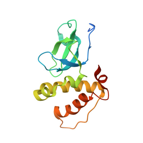 | |
UniProt & NIH Common Fund Data Resources | |||||
PHAROS: Q9UBC3 GTEx: ENSG00000088305 | |||||
Entity Groups | |||||
| Sequence Clusters | 30% Identity50% Identity70% Identity90% Identity95% Identity100% Identity | ||||
| UniProt Group | Q9UBC3 | ||||
Sequence AnnotationsExpand | |||||
Reference Sequence | |||||
| Length ( Å ) | Angle ( ˚ ) |
|---|---|
| a = 54.17 | α = 90 |
| b = 75.062 | β = 90 |
| c = 34.488 | γ = 90 |
| Software Name | Purpose |
|---|---|
| DENZO | data reduction |
| SCALEPACK | data scaling |
| REFMAC | refinement |
| PDB_EXTRACT | data extraction |


