Insights into pilus assembly and secretion from the structure and functional characterization of usher PapC.
Huang, Y., Smith, B.S., Chen, L.X., Baxter, R.H., Deisenhofer, J.(2009) Proc Natl Acad Sci U S A 106: 7403-7407
- PubMed: 19380723 Search on PubMedSearch on PubMed Central
- DOI: https://doi.org/10.1073/pnas.0902789106
- Primary Citation Related Structures:
3FIP - PubMed Abstract:
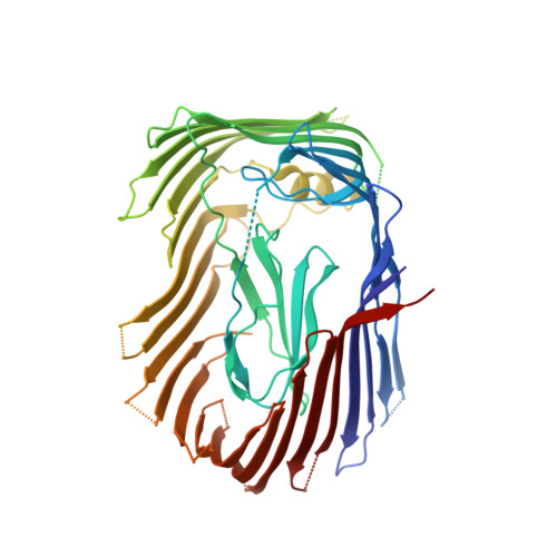
Ushers constitute a family of bacterial outer membrane proteins responsible for the assembly and secretion of surface organelles such as the pilus. The structure at 3.15-A resolution of the usher pyelonephritis-associated pili C (PapC) translocation domain reveals a 24-stranded kidney-shaped beta-barrel, occluded by an internal plug domain. The dimension of the pore allows tandem passage of individual folded pilus subunits in an upright pilus growth orientation, but is insufficient for accommodating donor strand exchange. The molecular packing revealed by the crystal structure shows that 2 PapC molecules in head-to-head orientation interact via exposed beta-strand edges, which could be the preferred dimer interaction in solution. In vitro reconstitution of fiber assemblies suggest that PapC monomers may be sufficient for fiber assembly and secretion; both the plug domain and the C-terminal domain of PapC are required for filament assembly, whereas the N-terminal domain is mainly responsible for recruiting the chaperone-subunit complexes to the usher. The plug domain has a dual function: gating the beta-pore and participating in pilus assembly.
- Howard Hughes Medical Institute and Department of Biochemistry, University of Texas Southwestern Medical Center, 6001 Forest Park Road, Dallas, TX 75390, USA. yihua.huang@utsouthwestern.edu
Organizational Affiliation: