


Crystal structure of the CBS-domain containing protein ATU1752 from Agrobacterium tumefaciens
Singer, A.U., Brown, G., Proudfoot, M., Xu, X., Dong, A., Cui, H., Edwards, A.M., Joachimiak, A., Savchenko, A., Yakunin, A.F.To be published.
Experimental Data Snapshot
Starting Model: experimental
View more details
Entity ID: 1 | |||||
|---|---|---|---|---|---|
| Molecule | Chains | Sequence Length | Organism | Details | Image |
| uncharacterized protein ATU1752 | 165 | Agrobacterium fabrum str. C58 | Mutation(s): 0 Gene Names: AGR_C_3216, Atu1752 | 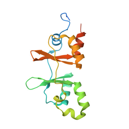 | |
UniProt | |||||
Entity Groups | |||||
| Sequence Clusters | 30% Identity50% Identity70% Identity90% Identity95% Identity100% Identity | ||||
| UniProt Group | A9CIP4 | ||||
Sequence AnnotationsExpand | |||||
Reference Sequence | |||||
| Ligands 3 Unique | |||||
|---|---|---|---|---|---|
| ID | Chains | Name / Formula / InChI Key | 2D Diagram | 3D Interactions | |
| NAI Download:Ideal Coordinates CCD File | F [auth A], I [auth B], L [auth C], O [auth D] | 1,4-DIHYDRONICOTINAMIDE ADENINE DINUCLEOTIDE C21 H29 N7 O14 P2 BOPGDPNILDQYTO-NNYOXOHSSA-N |  | ||
| AMP Download:Ideal Coordinates CCD File | E [auth A], H [auth B], K [auth C], N [auth D] | ADENOSINE MONOPHOSPHATE C10 H14 N5 O7 P UDMBCSSLTHHNCD-KQYNXXCUSA-N |  | ||
| SO4 Download:Ideal Coordinates CCD File | G [auth A], J [auth B], M [auth C], P [auth D] | SULFATE ION O4 S QAOWNCQODCNURD-UHFFFAOYSA-L |  | ||
| Length ( Å ) | Angle ( ˚ ) |
|---|---|
| a = 53.247 | α = 115.21 |
| b = 58.075 | β = 106.15 |
| c = 59.056 | γ = 94.94 |
| Software Name | Purpose |
|---|---|
| HKL-3000 | data collection |
| MrBUMP | phasing |
| REFMAC | refinement |
| HKL-3000 | data reduction |
| HKL-3000 | data scaling |


