3FD5 | pdb_00003fd5

Crystal structure of human selenophosphate synthetase 1 complex with AMPCP


Experimental Data Snapshot

  • Method: X-RAY DIFFRACTION
  • Resolution: 1.90 Å
  • R-Value Free: 
    0.195 (Depositor), 0.196 (DCC) 
  • R-Value Work: 
    0.138 (Depositor), 0.141 (DCC) 
  • R-Value Observed: 
    0.140 (Depositor) 

wwPDB Validation 3D Report Full Report

Validation slider image for 3FD5

Ligand Structure Quality Assessment 


This is version 1.2 of the entry. See complete history

Literature

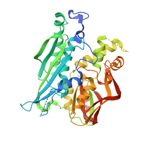
Crystal structures of catalytic intermediates of human selenophosphate synthetase 1.

Wang, K.T.Wang, J.Li, L.F.Su, X.D.

(2009) J Mol Biology 390: 747-759

  • DOI: https://doi.org/10.1016/j.jmb.2009.05.032
  • Primary Citation Related Structures: 
    3FD5, 3FD6

  • PubMed Abstract: 

    Selenophosphate synthetase catalyzes the synthesis of the highly active selenium donor molecule selenophosphate, a key intermediate in selenium metabolism. We have determined the high-resolution crystal structure of human selenophosphate synthetase 1 (hSPS1). An unexpected reaction intermediate, with a tightly bound phosphate and ADP at the active site has been captured in the structure. An enzymatic assay revealed that hSPS1 possesses low ADP hydrolysis activity in the presence of phosphate. Our structural and enzymatic results suggest that consuming the second high-energy phosphoester bond of ATP could protect the labile product selenophosphate during catalytic reaction. We solved another hSPS1 structure with potassium ions at the active sites. Comparing the two structures, we were able to define the monovalent cation-binding site of the enzyme. The detailed mechanism of the ADP hydrolysis step and the exact function of the monovalent cation for hSPS1 catalytic reaction are proposed.


  • Organizational Affiliation
    • National Laboratory of Protein Engineering and Plant Genetic Engineering, College of Life Sciences, Peking University, Beijing, P.R. China.

Macromolecule Content 

  • Total Structure Weight: 87.46 kDa 
  • Atom Count: 5,697 
  • Modeled Residue Count: 694 
  • Deposited Residue Count: 788 
  • Unique protein chains: 1

Macromolecules

Find similar proteins by:|  3D Structure
Entity ID: 1
MoleculeChains  Sequence LengthOrganismDetailsImage
Selenide, water dikinase 1
A, B
394Homo sapiensMutation(s): 0 
Gene Names: SEPHS1SELDSPSSPS1
EC: 2.7.9.3
UniProt & NIH Common Fund Data Resources
Find proteins for P49903 (Homo sapiens)
Explore P49903 
Go to UniProtKB:  P49903
PHAROS:  P49903
GTEx:  ENSG00000086475 
Entity Groups
Sequence Clusters30% Identity50% Identity70% Identity90% Identity95% Identity100% Identity
UniProt GroupP49903
Sequence Annotations
Expand
Reference Sequence

Small Molecules

Ligands 4 Unique
IDChains Name / Formula / InChI Key2D Diagram3D Interactions
AP2

Query on AP2


Download:Ideal Coordinates CCD File
C [auth A],
I [auth B]
PHOSPHOMETHYLPHOSPHONIC ACID ADENOSYL ESTER
C11 H17 N5 O9 P2
OLCWZBFDIYXLAA-IOSLPCCCSA-N
PO4

Query on PO4


Download:Ideal Coordinates CCD File
D [auth A],
J [auth B]
PHOSPHATE ION
O4 P
NBIIXXVUZAFLBC-UHFFFAOYSA-K
K

Query on K


Download:Ideal Coordinates CCD File
H [auth A],
M [auth B]
POTASSIUM ION
K
NPYPAHLBTDXSSS-UHFFFAOYSA-N
MG

Query on MG


Download:Ideal Coordinates CCD File
E [auth A]
F [auth A]
G [auth A]
K [auth B]
L [auth B]
E [auth A],
F [auth A],
G [auth A],
K [auth B],
L [auth B],
N [auth B]
MAGNESIUM ION
Mg
JLVVSXFLKOJNIY-UHFFFAOYSA-N

Experimental Data & Validation

Experimental Data

  • Method: X-RAY DIFFRACTION
  • Resolution: 1.90 Å
  • R-Value Free:  0.195 (Depositor), 0.196 (DCC) 
  • R-Value Work:  0.138 (Depositor), 0.141 (DCC) 
  • R-Value Observed: 0.140 (Depositor) 
Space Group: P 43
Unit Cell:
Length ( Å )Angle ( ˚ )
a = 66.734α = 90
b = 66.734β = 90
c = 181.168γ = 90
Software Package:
Software NamePurpose
HKL-2000data collection
PHENIXmodel building
PHENIXrefinement
HKL-2000data reduction
HKL-2000data scaling
PHENIXphasing

Structure Validation

View Full Validation Report


Ligand Structure Quality Assessment 


Entry History 

Deposition Data

  • Released Date: 2009-09-22 
  • Deposition Author(s): Wang, K.T.

Revision History  (Full details and data files)

  • Version 1.0: 2009-09-22
    Type: Initial release
  • Version 1.1: 2011-07-13
    Changes: Version format compliance
  • Version 1.2: 2023-12-27
    Changes: Data collection, Database references, Derived calculations