3FCU | pdb_00003fcu

Structure of headpiece of integrin aIIBb3 in open conformation


Experimental Data Snapshot

  • Method: X-RAY DIFFRACTION
  • Resolution: 2.90 Å
  • R-Value Free: 
    0.197 (Depositor), 0.197 (DCC) 
  • R-Value Work: 
    0.174 (Depositor), 0.177 (DCC) 
  • R-Value Observed: 
    0.174 (Depositor) 

Starting Model: experimental
View more details

wwPDB Validation 3D Report Full Report

Validation slider image for 3FCU

Ligand Structure Quality Assessment 


This is version 2.2 of the entry. See complete history

Literature

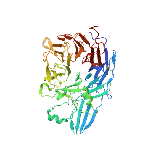
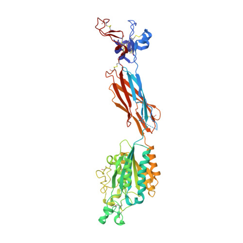
Structure of a complete integrin ectodomain in a physiologic resting state and activation and deactivation by applied forces.

Zhu, J.Luo, B.H.Xiao, T.Zhang, C.Nishida, N.Springer, T.A.

(2008) Mol Cell 32: 849-861

  • DOI: https://doi.org/10.1016/j.molcel.2008.11.018
  • Primary Citation Related Structures: 
    3FCS, 3FCU

  • PubMed Abstract: 

    The complete ectodomain of integrin alpha(IIb)beta(3) reveals a bent, closed, low-affinity conformation, the beta knee, and a mechanism for linking cytoskeleton attachment to high affinity for ligand. Ca and Mg ions in the recognition site, including the synergistic metal ion binding site (SyMBS), are loaded prior to ligand binding. Electrophilicity of the ligand-binding Mg ion is increased in the open conformation. The beta(3) knee passes between the beta(3)-PSI and alpha(IIb)-knob to bury the lower beta leg in a cleft, from which it is released for extension. Different integrin molecules in crystals and EM reveal breathing that appears on pathway to extension. Tensile force applied to the extended ligand-receptor complex stabilizes the closed, low-affinity conformation. By contrast, an additional lateral force applied to the beta subunit to mimic attachment to moving actin filaments stabilizes the open, high-affinity conformation. This mechanism propagates allostery over long distances and couples cytoskeleton attachment of integrins to their high-affinity state.


  • Organizational Affiliation
    • The Immune Disease Institute and Department of Pathology, Harvard Medical School, 200 Longwood Avenue, Boston, MA 02115, USA.

Macromolecule Content 

  • Total Structure Weight: 307.08 kDa 
  • Atom Count: 21,657 
  • Modeled Residue Count: 2,725 
  • Deposited Residue Count: 2,754 
  • Unique protein chains: 2

Macromolecules

Find similar proteins by:|  3D Structure
Entity ID: 1
MoleculeChains  Sequence LengthOrganismDetailsImage
Integrin, alpha 2b
A, C, E
457Homo sapiensMutation(s): 0 
Gene Names: ITGA2B
UniProt & NIH Common Fund Data Resources
Find proteins for P08514 (Homo sapiens)
Explore P08514 
Go to UniProtKB:  P08514
PHAROS:  P08514
GTEx:  ENSG00000005961 
Entity Groups
Sequence Clusters30% Identity50% Identity70% Identity90% Identity95% Identity100% Identity
UniProt GroupP08514
Glycosylation
Glycosylation Sites: 1Go to GlyGen: P08514-1
Sequence Annotations
Expand
Reference Sequence
Find similar proteins by:|  3D Structure
Entity ID: 2
MoleculeChains  Sequence LengthOrganismDetailsImage
Integrin beta-3
B, D, F
461Homo sapiensMutation(s): 0 
Gene Names: ITGB3GP3A
UniProt & NIH Common Fund Data Resources
Find proteins for P05106 (Homo sapiens)
Explore P05106 
Go to UniProtKB:  P05106
PHAROS:  P05106
GTEx:  ENSG00000259207 
Entity Groups
Sequence Clusters30% Identity50% Identity70% Identity90% Identity95% Identity100% Identity
UniProt GroupP05106
Glycosylation
Glycosylation Sites: 2Go to GlyGen: P05106-1
Sequence Annotations
Expand
Reference Sequence

Oligosaccharides

Help  
Entity ID: 3
MoleculeChains Length2D Diagram GlycosylationD Interactions
alpha-D-mannopyranose-(1-3)-[alpha-D-mannopyranose-(1-6)]alpha-D-mannopyranose-(1-4)-2-acetamido-2-deoxy-beta-D-glucopyranose-(1-4)-2-acetamido-2-deoxy-beta-D-glucopyranose
G, I
5N-Glycosylation
Glycosylation Resources
GlyTouCan: G21381MC
GlyCosmos: G21381MC
GlyGen: G21381MC
Entity ID: 4
MoleculeChains Length2D Diagram GlycosylationD Interactions
alpha-D-mannopyranose-(1-3)-alpha-D-mannopyranose-(1-4)-2-acetamido-2-deoxy-beta-D-glucopyranose-(1-4)-2-acetamido-2-deoxy-beta-D-glucopyranose
H
4N-Glycosylation
Glycosylation Resources
GlyTouCan: G48068RF
GlyCosmos: G48068RF
GlyGen: G48068RF

Small Molecules

Ligands 4 Unique
IDChains Name / Formula / InChI Key2D Diagram3D Interactions
NAG

Query on NAG


Download:Ideal Coordinates CCD File
EA [auth D]
FA [auth D]
LA [auth E]
N [auth A]
QA [auth F]
EA [auth D],
FA [auth D],
LA [auth E],
N [auth A],
QA [auth F],
S [auth B],
T [auth B],
Z [auth C]
2-acetamido-2-deoxy-beta-D-glucopyranose
C8 H15 N O6
OVRNDRQMDRJTHS-FMDGEEDCSA-N
CAC

Query on CAC


Download:Ideal Coordinates CCD File
GA [auth D],
RA [auth F],
U [auth B]
CACODYLATE ION
C2 H6 As O2
OGGXGZAMXPVRFZ-UHFFFAOYSA-M
CA

Query on CA


Download:Ideal Coordinates CCD File
CA [auth D]
DA [auth D]
HA [auth E]
IA [auth E]
J [auth A]
CA [auth D],
DA [auth D],
HA [auth E],
IA [auth E],
J [auth A],
JA [auth E],
K [auth A],
KA [auth E],
L [auth A],
M [auth A],
OA [auth F],
PA [auth F],
Q [auth B],
R [auth B],
V [auth C],
W [auth C],
X [auth C],
Y [auth C]
CALCIUM ION
Ca
BHPQYMZQTOCNFJ-UHFFFAOYSA-N
MG

Query on MG


Download:Ideal Coordinates CCD File
AA [auth C]
BA [auth D]
MA [auth E]
NA [auth F]
O [auth A]
AA [auth C],
BA [auth D],
MA [auth E],
NA [auth F],
O [auth A],
P [auth B]
MAGNESIUM ION
Mg
JLVVSXFLKOJNIY-UHFFFAOYSA-N

Experimental Data & Validation

Experimental Data

  • Method: X-RAY DIFFRACTION
  • Resolution: 2.90 Å
  • R-Value Free:  0.197 (Depositor), 0.197 (DCC) 
  • R-Value Work:  0.174 (Depositor), 0.177 (DCC) 
  • R-Value Observed: 0.174 (Depositor) 
Space Group: P 62
Unit Cell:
Length ( Å )Angle ( ˚ )
a = 332.093α = 90
b = 332.093β = 90
c = 88.288γ = 120
Software Package:
Software NamePurpose
ADSCdata collection
AMoREphasing
REFMACrefinement
HKL-2000data reduction
SCALEPACKdata scaling

Structure Validation

View Full Validation Report


Ligand Structure Quality Assessment 


Entry History 

Deposition Data

Revision History  (Full details and data files)

  • Version 1.0: 2009-01-20
    Type: Initial release
  • Version 1.1: 2011-07-13
    Changes: Advisory, Version format compliance
  • Version 2.0: 2020-07-29
    Type: Remediation
    Reason: Carbohydrate remediation
    Changes: Advisory, Atomic model, Data collection, Derived calculations, Structure summary
  • Version 2.1: 2023-09-06
    Changes: Data collection, Database references, Refinement description, Structure summary
  • Version 2.2: 2024-11-27
    Changes: Structure summary