3FCC

CRYSTAL STRUCTURE OF DLTA PROTEIN IN COMPLEX WITH ATP and MAGNESIUM


Experimental Data Snapshot

  • Method: X-RAY DIFFRACTION
  • Resolution: 2.32 Å
  • R-Value Free: 0.286 
  • R-Value Work: 0.241 
  • R-Value Observed: 0.241 

wwPDB Validation   3D Report Full Report


Ligand Structure Quality Assessment 


This is version 1.2 of the entry. See complete history


Literature

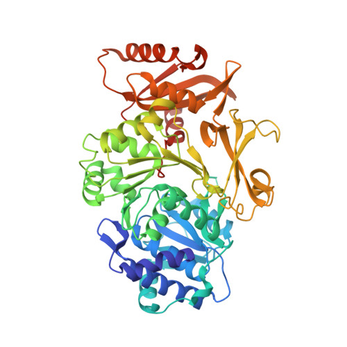
Crystal structure of Bacillus cereus D-alanyl carrier protein ligase (DltA) in complex with ATP.

Osman, K.T.Du, L.He, Y.Luo, Y.

(2009) J Mol Biol 388: 345-355

  • DOI: https://doi.org/10.1016/j.jmb.2009.03.040
  • Primary Citation of Related Structures:  
    3FCC, 3FCE

  • PubMed Abstract: 

    D-alanylation of lipoteichoic acids modulates the surface charge and ligand binding of the Gram-positive cell wall. Disruption of the bacterial dlt operon involved in teichoic acid alanylation, as well as inhibition of the DltA (D-alanyl carrier protein ligase) protein, has been shown to render the bacterium more susceptible to conventional antibiotics and host defense responses. The DltA catalyzes the adenylation and thiolation reactions of d-alanine. This enzyme belongs to a superfamily of AMP-forming domains such as the ubiquitous acetyl-coenzyme A synthetase. We have determined the 1.9-A-resolution crystal structure of a DltA protein from Bacillus cereus in complex with ATP. This structure sheds light on the geometry of the bound ATP. The invariant catalytic residue Lys492 appears to be mobile, suggesting a molecular mechanism of catalysis for this superfamily of enzymes. Specific roles are also revealed for two other invariant residues: the divalent cation-stabilizing Glu298 and the beta-phosphate-interacting Arg397. Mutant proteins with a glutamine substitution at position 298 or 397 are inactive.


  • Organizational Affiliation

    Department of Biochemistry, University of Saskatchewan, A3 Health Sciences Building, 107 Wiggins Road, Saskatoon, Saskatchewan, Canada S7N 5E5.


Macromolecules
Find similar proteins by:  (by identity cutoff)  |  3D Structure
Entity ID: 1
MoleculeChains Sequence LengthOrganismDetailsImage
D-alanine--poly(phosphoribitol) ligase subunit 1512Bacillus cereus ATCC 14579Mutation(s): 0 
Gene Names: BC_1372dltA
EC: 6.1.1.13
UniProt
Find proteins for Q81G39 (Bacillus cereus (strain ATCC 14579 / DSM 31 / CCUG 7414 / JCM 2152 / NBRC 15305 / NCIMB 9373 / NCTC 2599 / NRRL B-3711))
Explore Q81G39 
Go to UniProtKB:  Q81G39
Entity Groups  
Sequence Clusters30% Identity50% Identity70% Identity90% Identity95% Identity100% Identity
UniProt GroupQ81G39
Sequence Annotations
Expand
  • Reference Sequence
Small Molecules
Ligands 2 Unique
IDChains Name / Formula / InChI Key2D Diagram3D Interactions
ATP
Query on ATP

Download Ideal Coordinates CCD File 
C [auth A]ADENOSINE-5'-TRIPHOSPHATE
C10 H16 N5 O13 P3
ZKHQWZAMYRWXGA-KQYNXXCUSA-N
MG
Query on MG

Download Ideal Coordinates CCD File 
B [auth A]MAGNESIUM ION
Mg
JLVVSXFLKOJNIY-UHFFFAOYSA-N
Experimental Data & Validation

Experimental Data

  • Method: X-RAY DIFFRACTION
  • Resolution: 2.32 Å
  • R-Value Free: 0.286 
  • R-Value Work: 0.241 
  • R-Value Observed: 0.241 
  • Space Group: P 1 21 1
Unit Cell:
Length ( Å )Angle ( ˚ )
a = 61.6α = 90
b = 87β = 115
c = 60.1γ = 90
Software Package:
Software NamePurpose
XDSdata scaling
CNSrefinement
XDSdata reduction
CNSphasing

Structure Validation

View Full Validation Report


Ligand Structure Quality Assessment 


Entry History 

Deposition Data

Revision History  (Full details and data files)

  • Version 1.0: 2009-04-14
    Type: Initial release
  • Version 1.1: 2011-07-13
    Changes: Source and taxonomy, Version format compliance
  • Version 1.2: 2023-09-06
    Changes: Data collection, Database references, Derived calculations, Refinement description