


Oxygen Activation in Neuronal NO Synthase: Stabilisation of a Novel Intermediate in the G586S Mutant
Papale, D., Bruckmann, C., Miles, C.S., Mowat, C.G., Daff, S.To be published.
Experimental Data Snapshot
Starting Model: experimental
View more details
Entity ID: 1 | |||||
|---|---|---|---|---|---|
| Molecule | Chains | Sequence Length | Organism | Details | Image |
| Nitric oxide synthase, brain | 422 | Rattus norvegicus | Mutation(s): 1 Gene Names: NOS1 or BNOS EC: 1.14.13.39 | 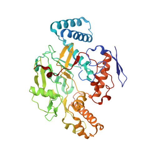 | |
UniProt | |||||
Entity Groups | |||||
| Sequence Clusters | 30% Identity50% Identity70% Identity90% Identity95% Identity100% Identity | ||||
| UniProt Group | P29476 | ||||
Sequence AnnotationsExpand | |||||
Reference Sequence | |||||
| Ligands 4 Unique | |||||
|---|---|---|---|---|---|
| ID | Chains | Name / Formula / InChI Key | 2D Diagram | 3D Interactions | |
| HEM Download:Ideal Coordinates CCD File | C [auth A], H [auth B] | PROTOPORPHYRIN IX CONTAINING FE C34 H32 Fe N4 O4 KABFMIBPWCXCRK-RGGAHWMASA-L |  | ||
| H4B Download:Ideal Coordinates CCD File | E [auth A], G [auth B] | 5,6,7,8-TETRAHYDROBIOPTERIN C9 H15 N5 O3 FNKQXYHWGSIFBK-RPDRRWSUSA-N |  | ||
| ARG Download:Ideal Coordinates CCD File | F [auth A], I [auth B] | ARGININE C6 H15 N4 O2 ODKSFYDXXFIFQN-BYPYZUCNSA-O |  | ||
| ZN Download:Ideal Coordinates CCD File | D [auth A] | ZINC ION Zn PTFCDOFLOPIGGS-UHFFFAOYSA-N |  | ||
| Length ( Å ) | Angle ( ˚ ) |
|---|---|
| a = 51.623 | α = 90 |
| b = 110.276 | β = 90 |
| c = 164.671 | γ = 90 |
| Software Name | Purpose |
|---|---|
| PHASER | phasing |
| PHENIX | refinement |
| MOSFLM | data reduction |
| SCALA | data scaling |


