3F7P

Crystal structure of a complex between integrin beta4 and plectin


Experimental Data Snapshot

  • Method: X-RAY DIFFRACTION
  • Resolution: 2.75 Å
  • R-Value Free: 0.257 
  • R-Value Work: 0.204 
  • R-Value Observed: 0.207 

wwPDB Validation   3D Report Full Report


This is version 1.2 of the entry. See complete history


Literature

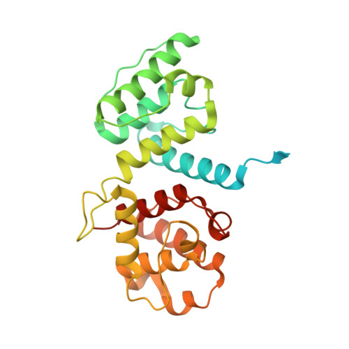
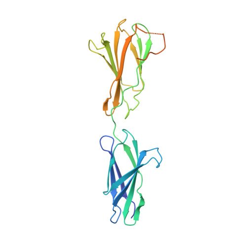
Structural basis of the interaction between integrin alpha6beta4 and plectin at the hemidesmosomes

de Pereda, J.M.Lillo, M.P.Sonnenberg, A.

(2009) EMBO J 28: 1180-1190

  • DOI: https://doi.org/10.1038/emboj.2009.48
  • Primary Citation of Related Structures:  
    3F7P, 3F7Q, 3F7R

  • PubMed Abstract: 

    The interaction between the integrin alpha6beta4 and plectin is essential for the assembly and stability of hemidesmosomes, which are junctional adhesion complexes that anchor epithelial cells to the basement membrane. We describe the crystal structure at 2.75 A resolution of the primary alpha6beta4-plectin complex, formed by the first pair of fibronectin type III domains and the N-terminal region of the connecting segment of beta4 and the actin-binding domain of plectin. Two missense mutations in beta4 (R1225H and R1281W) linked to nonlethal forms of epidermolysis bullosa prevent essential intermolecular contacts. We also present two structures at 1.75 and 2.05 A resolution of the beta4 moiety in the absence of plectin, which reveal a major rearrangement of the connecting segment of beta4 on binding to plectin. This conformational switch is correlated with the way alpha6beta4 promotes stable adhesion or cell migration and suggests an allosteric control of the integrin.


  • Organizational Affiliation

    Department of Structural Biology, Instituto de Biología Molecular y Celular del Cáncer, Consejo Superior de Investigaciones Científicas-Universidad de Salamanca, Campus Unamuno, Salamanca, Spain. pereda@usal.es


Macromolecules
Find similar proteins by:  (by identity cutoff)  |  3D Structure
Entity ID: 1
MoleculeChains Sequence LengthOrganismDetailsImage
Plectin-1
A, B
296Homo sapiensMutation(s): 0 
Gene Names: PLEC-1 (Isoform 1C)PLEC1
UniProt & NIH Common Fund Data Resources
Find proteins for Q15149 (Homo sapiens)
Explore Q15149 
Go to UniProtKB:  Q15149
PHAROS:  Q15149
GTEx:  ENSG00000178209 
Entity Groups  
Sequence Clusters30% Identity50% Identity70% Identity90% Identity95% Identity100% Identity
UniProt GroupQ15149
Sequence Annotations
Expand
  • Reference Sequence
Find similar proteins by:  (by identity cutoff)  |  3D Structure
Entity ID: 2
MoleculeChains Sequence LengthOrganismDetailsImage
Integrin beta-4
C, D, E
248Homo sapiensMutation(s): 0 
Gene Names: ITGB4
UniProt & NIH Common Fund Data Resources
Find proteins for P16144 (Homo sapiens)
Explore P16144 
Go to UniProtKB:  P16144
PHAROS:  P16144
GTEx:  ENSG00000132470 
Entity Groups  
Sequence Clusters30% Identity50% Identity70% Identity90% Identity95% Identity100% Identity
UniProt GroupP16144
Sequence Annotations
Expand
  • Reference Sequence
Experimental Data & Validation

Experimental Data

  • Method: X-RAY DIFFRACTION
  • Resolution: 2.75 Å
  • R-Value Free: 0.257 
  • R-Value Work: 0.204 
  • R-Value Observed: 0.207 
  • Space Group: P 32 2 1
Unit Cell:
Length ( Å )Angle ( ˚ )
a = 107.25α = 90
b = 107.25β = 90
c = 203.97γ = 120
Software Package:
Software NamePurpose
DNAdata collection
PHASERphasing
PHENIXrefinement
XDSdata reduction
XSCALEdata scaling

Structure Validation

View Full Validation Report


Entry History 

Deposition Data

Revision History  (Full details and data files)

  • Version 1.0: 2009-03-10
    Type: Initial release
  • Version 1.1: 2011-07-13
    Changes: Version format compliance
  • Version 1.2: 2023-11-01
    Changes: Data collection, Database references, Derived calculations, Refinement description