3F74 | pdb_00003f74

Crystal structure of wild type LFA1 I domain


Experimental Data Snapshot

  • Method: X-RAY DIFFRACTION
  • Resolution: 1.70 Å
  • R-Value Free: 
    0.178 (Depositor), 0.213 (DCC) 
  • R-Value Work: 
    0.146 (Depositor) 
  • R-Value Observed: 
    0.147 (Depositor) 

wwPDB Validation 3D Report Full Report

Validation slider image for 3F74

This is version 1.3 of the entry. See complete history

Literature

Crystal structure of isoflurane bound to integrin LFA-1 supports a unified mechanism of volatile anesthetic action in the immune and central nervous systems.

Zhang, H.Astrof, N.S.Liu, J.H.Wang, J.H.Shimaoka, M.

(2009) FASEB J 23: 2735-2740

  • DOI: https://doi.org/10.1096/fj.09-129908
  • Primary Citation Related Structures: 
    3F74, 3F78

  • PubMed Abstract: 

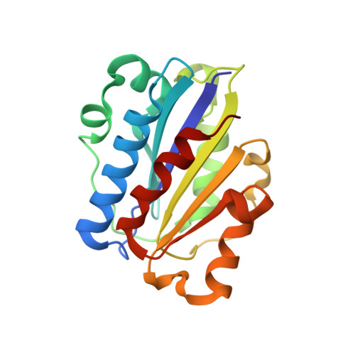
    Volatile anesthetics (VAs), such as isoflurane, induce a general anesthetic state by binding to specific targets (i.e., ion channels) in the central nervous system (CNS). Simultaneously, VAs modulate immune functions, possibly via direct interaction with alternative targets on leukocytes. One such target, the integrin lymphocyte function-associated antigen-1 (LFA-1), has been shown previously to be inhibited by isoflurane. A better understanding of the mechanism by which isoflurane alters protein function requires the detailed information about the drug-protein interaction at an atomic level. Here, we describe the crystal structure of the LFA-1 ligand-binding domain (I domain) in complex with isoflurane at 1.6 A. We discovered that isoflurane binds to an allosteric cavity previously implicated as critical for the transition of LFA-1 from the low- to the high-affinity state. The isoflurane binding site in the I domain involves an array of amphiphilic interactions, thereby resembling a "common anesthetic binding motif" previously predicted for authentic VA binding sites. These results suggest that the allosteric modulation of protein function by isoflurane, as demonstrated for the integrin LFA-1, might represent a unified mechanism shared by the interactions of volatile anesthetics with targets in the CNS.


  • Organizational Affiliation
    • Dana-Farber Cancer Institute, Boston, Massachusetts, USA.

Macromolecule Content 

  • Total Structure Weight: 62.43 kDa 
  • Atom Count: 5,147 
  • Modeled Residue Count: 540 
  • Deposited Residue Count: 543 
  • Unique protein chains: 1

Macromolecules

Find similar proteins by:|  3D Structure
Entity ID: 1
MoleculeChains  Sequence LengthOrganismDetailsImage
Integrin alpha-L
A, B, C
181Homo sapiensMutation(s): 1 
Gene Names: CD11Aintegrin LFA1ITGAL
UniProt & NIH Common Fund Data Resources
Find proteins for P20701 (Homo sapiens)
Explore P20701 
Go to UniProtKB:  P20701
PHAROS:  P20701
GTEx:  ENSG00000005844 
Entity Groups
Sequence Clusters30% Identity50% Identity70% Identity90% Identity95% Identity100% Identity
UniProt GroupP20701
Sequence Annotations
Expand
Reference Sequence

Experimental Data & Validation

Experimental Data

  • Method: X-RAY DIFFRACTION
  • Resolution: 1.70 Å
  • R-Value Free:  0.178 (Depositor), 0.213 (DCC) 
  • R-Value Work:  0.146 (Depositor) 
  • R-Value Observed: 0.147 (Depositor) 
Space Group: P 1 21 1
Unit Cell:
Length ( Å )Angle ( ˚ )
a = 61.806α = 90
b = 85.603β = 117.93
c = 63.554γ = 90
Software Package:
Software NamePurpose
DENZOdata reduction
SCALEPACKdata scaling
REFMACrefinement
PDB_EXTRACTdata extraction
HKL-3000data collection
PHASERphasing

Structure Validation

View Full Validation Report


Entry History 

Deposition Data

Revision History  (Full details and data files)

  • Version 1.0: 2009-06-23
    Type: Initial release
  • Version 1.1: 2011-07-13
    Changes: Advisory, Version format compliance
  • Version 1.2: 2017-10-25
    Changes: Refinement description
  • Version 1.3: 2023-12-27
    Changes: Data collection, Database references, Derived calculations