3EZW

Crystal Structure of a Hyperactive Escherichia coli Glycerol Kinase Mutant Gly230 --> Asp Obtained Using Microfluidic Crystallization Devices


Experimental Data Snapshot

  • Method: X-RAY DIFFRACTION
  • Resolution: 2.00 Å
  • R-Value Free: 0.225 
  • R-Value Work: 0.167 
  • R-Value Observed: 0.168 

wwPDB Validation   3D Report Full Report


This is version 1.4 of the entry. See complete history


Literature

Crystal structure of a hyperactive Escherichia coli glycerol kinase mutant Gly230 --> Asp obtained using microfluidic crystallization devices.

Anderson, M.J.DeLabarre, B.Raghunathan, A.Palsson, B.O.Brunger, A.T.Quake, S.R.

(2007) Biochemistry 46: 5722-5731

  • DOI: https://doi.org/10.1021/bi700096p
  • Primary Citation of Related Structures:  
    3EZW

  • PubMed Abstract: 

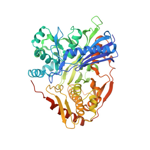
    The crystal structure of an Escherichia coli glycerol kinase mutant Gly230 --> Asp (GKG230D) was determined to 2.0 A resolution using a microfluidics based crystallization platform. The crystallization strategy involved a suite of microfluidic devices that characterized the solubility trends of GKG230D, performed nanoliter volume free interface diffusion crystallization experiments, and produced diffraction-quality crystals for in situ data collection. GKG230D displays increased enzymatic activity and decreased allosteric regulation by the glycolytic pathway intermediate fructose 1,6-bisphosphate (FBP) compared to wild-type GK (GKWT). Structural analysis revealed that the decreased allosteric regulation is a result of the altered FBP binding loop conformations in GKG230D that interfere with the wild-type FBP binding site. The altered FBP binding loop conformations in GKG230D are supported through a series of intramolecular loop interactions. The appearance of Asp230 in the FBP binding loops also repositions the wild-type FBP binding residues away from the FBP binding site. Light scattering analysis confirmed GKG230D is a dimer and is resistant to tetramer formation in the presence of FBP, whereas GKWT dimers are converted into putatively inactive tetramers in the presence of FBP. GKG230D also provides the first structural evidence for multiple GK monomer conformations in the presence of glycerol and in the absence of a nucleotide substrate and verifies that glycerol binding is not responsible for locking GK into the closed conformation necessary for GK activity.


  • Organizational Affiliation

    Department of Biochemistry and Molecular Biophysics, California Institute of Technology, MS 128-95, Pasadena, California 91125, USA.


Macromolecules
Find similar proteins by:  (by identity cutoff)  |  3D Structure
Entity ID: 1
MoleculeChains Sequence LengthOrganismDetailsImage
Glycerol kinase526Escherichia coli K-12Mutation(s): 1 
Gene Names: b3926glpKJW3897
EC: 2.7.1.30
UniProt
Find proteins for P0A6F3 (Escherichia coli (strain K12))
Explore P0A6F3 
Go to UniProtKB:  P0A6F3
Entity Groups  
Sequence Clusters30% Identity50% Identity70% Identity90% Identity95% Identity100% Identity
UniProt GroupP0A6F3
Sequence Annotations
Expand
  • Reference Sequence
Small Molecules
Ligands 4 Unique
IDChains Name / Formula / InChI Key2D Diagram3D Interactions
GOL
Query on GOL

Download Ideal Coordinates CCD File 
AA [auth C]
GA [auth D]
I [auth A]
KA [auth F]
N [auth B]
AA [auth C],
GA [auth D],
I [auth A],
KA [auth F],
N [auth B],
OA [auth H],
R [auth E],
U [auth G]
GLYCEROL
C3 H8 O3
PEDCQBHIVMGVHV-UHFFFAOYSA-N
EDO
Query on EDO

Download Ideal Coordinates CCD File 
CA [auth C]
DA [auth C]
EA [auth C]
FA [auth C]
IA [auth D]
CA [auth C],
DA [auth C],
EA [auth C],
FA [auth C],
IA [auth D],
JA [auth D],
L [auth A],
M [auth A],
MA [auth F],
NA [auth F],
P [auth B],
Q [auth B],
QA [auth H],
RA [auth H],
T [auth E],
W [auth G],
X [auth G],
Y [auth G],
Z [auth G]
1,2-ETHANEDIOL
C2 H6 O2
LYCAIKOWRPUZTN-UHFFFAOYSA-N
CL
Query on CL

Download Ideal Coordinates CCD File 
BA [auth C]
HA [auth D]
J [auth A]
LA [auth F]
O [auth B]
BA [auth C],
HA [auth D],
J [auth A],
LA [auth F],
O [auth B],
PA [auth H],
S [auth E],
V [auth G]
CHLORIDE ION
Cl
VEXZGXHMUGYJMC-UHFFFAOYSA-M
MG
Query on MG

Download Ideal Coordinates CCD File 
K [auth A]MAGNESIUM ION
Mg
JLVVSXFLKOJNIY-UHFFFAOYSA-N
Experimental Data & Validation

Experimental Data

  • Method: X-RAY DIFFRACTION
  • Resolution: 2.00 Å
  • R-Value Free: 0.225 
  • R-Value Work: 0.167 
  • R-Value Observed: 0.168 
  • Space Group: P 1 21 1
Unit Cell:
Length ( Å )Angle ( ˚ )
a = 91.116α = 90
b = 114.26β = 91.15
c = 212.624γ = 90
Software Package:
Software NamePurpose
REFMACrefinement
CNSrefinement
DENZOdata reduction
SCALEPACKdata scaling
CNSphasing

Structure Validation

View Full Validation Report


Entry History 

Deposition Data

Revision History  (Full details and data files)

  • Version 1.0: 2008-11-04
    Type: Initial release
  • Version 1.1: 2011-07-13
    Changes: Non-polymer description, Version format compliance
  • Version 1.2: 2019-07-24
    Changes: Data collection, Refinement description
  • Version 1.3: 2021-10-20
    Changes: Database references, Derived calculations
  • Version 1.4: 2023-09-06
    Changes: Data collection, Refinement description