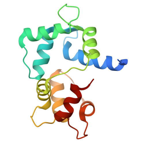
3EWV

Crystal Structure of calmodulin complexed with a peptide


Experimental Data Snapshot

  • Method: X-RAY DIFFRACTION
  • Resolution: 2.60 Å
  • R-Value Free: 0.282 
  • R-Value Work: 0.228 
  • R-Value Observed: 0.231 

Starting Model: experimental
View more details

wwPDB Validation   3D Report Full Report


This is version 1.4 of the entry. See complete history


Literature

Structural insights into the mechanism of calmodulin binding to death receptors.

Cao, P.Zhang, W.Gui, W.Dong, Y.Jiang, T.Gong, Y.

(2014) Acta Crystallogr D Biol Crystallogr 70: 1604-1613

  • DOI: https://doi.org/10.1107/S1399004714006919
  • Primary Citation of Related Structures:  
    3EWT, 3EWV

  • PubMed Abstract: 

    The death receptors Fas, p75(NTR) and DR6 are key components of extrinsically activated apoptosis. Characterization of how they interact with the adaptors is crucial in order to unravel the signalling mechanisms. However, the exact conformation that their intracellular death domain adopts upon binding downstream partners remains unclear. One model suggests that it adopts a typical compact fold, whilst a second model proposed an open conformation. Calmodulin (CaM), a major calcium sensor, has previously been reported to be one of the Fas adaptors that modulate apoptosis. This work reports that CaM also binds directly to the death domains of p75(NTR) and DR6, indicating that it serves as a common modulator of the death receptors. Two crystal structures of CaM in complexes with the corresponding binding regions of Fas and p75(NTR) are also reported. Interestingly, the precise CaM-binding sites were mapped to different regions: helix 1 in Fas and helix 5 in p75(NTR) and DR6. A novel 1-11 motif for CaM binding was observed in p75(NTR). Modelling the complexes of CaM with full-length receptors reveals that the opening of the death domains would be essential in order to expose their binding sites for CaM. These results may facilitate understanding of the diverse functional repertoire of death receptors and CaM and provide further insights necessary for the design of potential therapeutic peptide agents.


  • Organizational Affiliation

    National Laboratory of Biomacromolecules, Institute of Biophysics, Chinese Academy of Sciences, 15 Datun Road, Chaoyang District, Beijing 100101, People's Republic of China.


Macromolecules
Find similar proteins by:  (by identity cutoff)  |  3D Structure
Entity ID: 1
MoleculeChains Sequence LengthOrganismDetailsImage
Calmodulin154Homo sapiensMutation(s): 0 
UniProt & NIH Common Fund Data Resources
Find proteins for P0DP23 (Homo sapiens)
Explore P0DP23 
Go to UniProtKB:  P0DP23
PHAROS:  P0DP23
GTEx:  ENSG00000198668 
Entity Groups  
Sequence Clusters30% Identity50% Identity70% Identity90% Identity95% Identity100% Identity
UniProt GroupP0DP23
Sequence Annotations
Expand
  • Reference Sequence

Find similar proteins by:  Sequence   |   3D Structure  

Entity ID: 2
MoleculeChains Sequence LengthOrganismDetailsImage
Tumor necrosis factor receptor superfamily member 16B [auth E]17synthetic constructMutation(s): 0 
UniProt & NIH Common Fund Data Resources
Find proteins for P08138 (Homo sapiens)
Explore P08138 
Go to UniProtKB:  P08138
PHAROS:  P08138
GTEx:  ENSG00000064300 
Entity Groups  
Sequence Clusters30% Identity50% Identity70% Identity90% Identity95% Identity100% Identity
UniProt GroupP08138
Sequence Annotations
Expand
  • Reference Sequence
Experimental Data & Validation

Experimental Data

  • Method: X-RAY DIFFRACTION
  • Resolution: 2.60 Å
  • R-Value Free: 0.282 
  • R-Value Work: 0.228 
  • R-Value Observed: 0.231 
  • Space Group: P 32 2 1
Unit Cell:
Length ( Å )Angle ( ˚ )
a = 39.936α = 90
b = 39.936β = 90
c = 181.993γ = 120
Software Package:
Software NamePurpose
CrystalCleardata collection
MOLREPphasing
REFMACrefinement
DENZOdata reduction
SCALEPACKdata scaling

Structure Validation

View Full Validation Report


Entry History 

Deposition Data

Revision History  (Full details and data files)

  • Version 1.0: 2009-10-20
    Type: Initial release
  • Version 1.1: 2011-07-13
    Changes: Version format compliance
  • Version 1.2: 2014-06-18
    Changes: Database references
  • Version 1.3: 2020-01-01
    Changes: Database references, Source and taxonomy
  • Version 1.4: 2023-11-01
    Changes: Data collection, Database references, Derived calculations, Refinement description