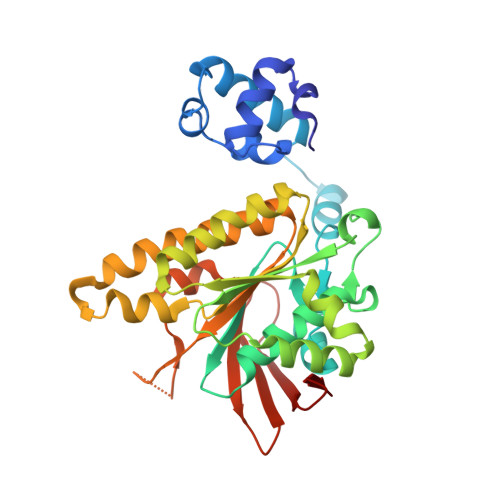
Conservation of a conformational switch in RadA recombinase from Methanococcus maripaludis.
Li, Y., He, Y., Luo, Y.(2009) Acta Crystallogr D Biol Crystallogr 65: 602-610
- PubMed: 19465774 Search on PubMedSearch on PubMed Central
- DOI: https://doi.org/10.1107/S0907444909011871
- Primary Citation Related Structures:
3ETL, 3EW9, 3EWA - PubMed Abstract:
Archaeal RadAs are close homologues of eukaryal Rad51s ( approximately 40% sequence identity). These recombinases promote ATP hydrolysis and a hallmark strand-exchange reaction between homologous single-stranded and double-stranded DNA substrates. Pairing of the 3'-overhangs located at the damaged DNA with a homologous double-stranded DNA enables the re-synthesis of the damaged region using the homologous DNA as the template. In recent studies, conformational changes in the DNA-interacting regions of Methanococcus voltae RadA have been correlated with the presence of activity-stimulating potassium or calcium ions in the ATPase centre. The series of crystal structures of M. maripaludis RadA presented here further suggest the conservation of an allosteric switch in the ATPase centre which controls the conformational status of DNA-interacting loops. Structural comparison with the distant Escherichia coli RecA homologue supports the notion that the conserved Lys248 and Lys250 residues in RecA play a role similar to that of cations in RadA. The conservation of a cationic bridge between the DNA-interacting L2 region and the terminal phosphate of ATP, together with the apparent stability of the nucleoprotein filament, suggests a gap-displacement model which may explain the advantage of ATP hydrolysis for DNA-strand exchange.
- Department of Biochemistry, University of Saskatchewan, Saskatoon, Saskatchewan S7N 5E5, Canada.
Organizational Affiliation: