3ETJ | pdb_00003etj

Crystal structure E. coli Purk in complex with Mg, ADP, and Pi


Experimental Data Snapshot

  • Method: X-RAY DIFFRACTION
  • Resolution: 1.60 Å
  • R-Value Free: 
    0.215 (Depositor) 
  • R-Value Work: 
    0.158 (Depositor) 
  • R-Value Observed: 
    0.160 (Depositor) 

Starting Model: experimental
View more details

wwPDB Validation 3D Report Full Report

Validation slider image for 3ETJ

Ligand Structure Quality Assessment 


This is version 1.3 of the entry. See complete history

Literature

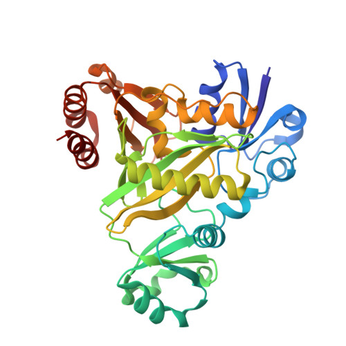
Structural analysis of the active site geometry of N(5)-Carboxyaminoimidazole ribonucleotide synthetase from Escherichia coli.

Thoden, J.B.Holden, H.M.Firestine, S.M.

(2008) Biochemistry 47: 13346-13353

  • DOI: https://doi.org/10.1021/bi801734z
  • Primary Citation Related Structures: 
    3ETH, 3ETJ

  • PubMed Abstract: 

    N(5)-Carboxyaminoimidazole ribonucleotide synthetase (N(5)-CAIR synthetase) converts 5-aminoimidazole ribonucleotide (AIR), MgATP, and bicarbonate into N(5)-CAIR, MgADP, and P(i). The enzyme is required for de novo purine biosynthesis in microbes yet is not found in humans suggesting that it represents an ideal and unexplored target for antimicrobial drug design. Here we report the X-ray structures of N(5)-CAIR synthetase from Escherichia coli with either MgATP or MgADP/P(i) bound in the active site cleft. These structures, determined to 1.6-A resolution, provide detailed information regarding the active site geometry before and after ATP hydrolysis. In both structures, two magnesium ions are observed. Each of these is octahedrally coordinated, and the carboxylate side chain of Glu238 bridges them. For the structure of the MgADP/P(i) complex, crystals were grown in the presence of AIR and MgATP. No electron density was observed for AIR, and the electron density corresponding to the nucleotide clearly revealed the presence of ADP and P(i) rather than ATP. The bound P(i) shifts by approximately 3 A relative to the gamma-phosphoryl group of ATP and forms electrostatic interactions with the side chains of Arg242 and His244. Since the reaction mechanism of N(5)-CAIR synthetase is believed to proceed via a carboxyphosphate intermediate, we propose that the location of the inorganic phosphate represents the binding site for stabilization of this reactive species. Using the information derived from the two structures reported here, coupled with molecular modeling, we propose a catalytic mechanism for N(5)-CAIR synthetase.


  • Organizational Affiliation
    • Department of Biochemistry, University of Wisconsin, Madison, Wisconsin 53706, USA.

Macromolecule Content 

  • Total Structure Weight: 80.24 kDa 
  • Atom Count: 6,305 
  • Modeled Residue Count: 706 
  • Deposited Residue Count: 710 
  • Unique protein chains: 1

Macromolecules

Find similar proteins by:|  3D Structure
Entity ID: 1
MoleculeChains  Sequence LengthOrganismDetailsImage
Phosphoribosylaminoimidazole carboxylase ATPase subunit
A, B
355Escherichia coli K-12Mutation(s): 2 
Gene Names: b0522JW0511purKPURK_ECOLI
EC: 4.1.1.21 (PDB Primary Data), 6.3.4.18 (UniProt)
UniProt
Find proteins for P09029 (Escherichia coli (strain K12))
Explore P09029 
Go to UniProtKB:  P09029
Entity Groups
Sequence Clusters30% Identity50% Identity70% Identity90% Identity95% Identity100% Identity
UniProt GroupP09029
Sequence Annotations
Expand
Reference Sequence

Small Molecules

Ligands 4 Unique
IDChains Name / Formula / InChI Key2D Diagram3D Interactions
ADP

Query on ADP


Download:Ideal Coordinates CCD File
C [auth A],
H [auth B]
ADENOSINE-5'-DIPHOSPHATE
C10 H15 N5 O10 P2
XTWYTFMLZFPYCI-KQYNXXCUSA-N
PI

Query on PI


Download:Ideal Coordinates CCD File
F [auth A],
K [auth B]
HYDROGENPHOSPHATE ION
H O4 P
NBIIXXVUZAFLBC-UHFFFAOYSA-L
CL

Query on CL


Download:Ideal Coordinates CCD File
G [auth A]CHLORIDE ION
Cl
VEXZGXHMUGYJMC-UHFFFAOYSA-M
MG

Query on MG


Download:Ideal Coordinates CCD File
D [auth A],
E [auth A],
I [auth B],
J [auth B]
MAGNESIUM ION
Mg
JLVVSXFLKOJNIY-UHFFFAOYSA-N

Experimental Data & Validation

Experimental Data

  • Method: X-RAY DIFFRACTION
  • Resolution: 1.60 Å
  • R-Value Free:  0.215 (Depositor) 
  • R-Value Work:  0.158 (Depositor) 
  • R-Value Observed: 0.160 (Depositor) 
Space Group: C 1 2 1
Unit Cell:
Length ( Å )Angle ( ˚ )
a = 128.8α = 90
b = 70.5β = 124.3
c = 88.7γ = 90
Software Package:
Software NamePurpose
TNTrefinement
PDB_EXTRACTdata extraction
HKL-2000data collection
HKL-2000data reduction
HKL-2000data scaling
PHASERphasing

Structure Validation

View Full Validation Report


Ligand Structure Quality Assessment 


Entry History 

Deposition Data

Revision History  (Full details and data files)

  • Version 1.0: 2008-10-21
    Type: Initial release
  • Version 1.1: 2011-07-13
    Changes: Version format compliance
  • Version 1.2: 2021-10-20
    Changes: Database references, Derived calculations
  • Version 1.3: 2023-09-06
    Changes: Data collection, Refinement description