3ETG | pdb_00003etg

Glutamate dehydrogenase complexed with GW5074


Experimental Data Snapshot

  • Method: X-RAY DIFFRACTION
  • Resolution: 2.50 Å
  • R-Value Free: 
    0.258 (Depositor), 0.254 (DCC) 
  • R-Value Work: 
    0.244 (Depositor), 0.238 (DCC) 
  • R-Value Observed: 
    0.244 (Depositor) 

wwPDB Validation 3D Report Full Report

Validation slider image for 3ETG

Ligand Structure Quality Assessment 


This is version 1.3 of the entry. See complete history

Literature

Novel Inhibitors Complexed with Glutamate Dehydrogenase: ALLOSTERIC REGULATION BY CONTROL OF PROTEIN DYNAMICS

Li, M.Smith, C.J.Walker, M.T.Smith, T.J.

(2009) J Biological Chem 284: 22988-23000

  • DOI: https://doi.org/10.1074/jbc.M109.020222
  • Primary Citation Related Structures: 
    3ETD, 3ETE, 3ETG

  • PubMed Abstract: 

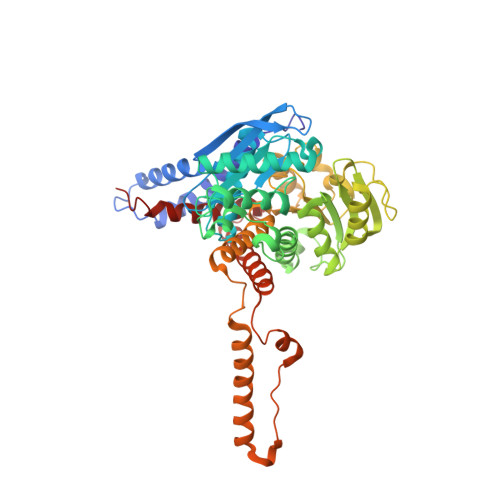
    Mammalian glutamate dehydrogenase (GDH) is a homohexameric enzyme that catalyzes the reversible oxidative deamination of l-glutamate to 2-oxoglutarate using NAD(P)(+) as coenzyme. Unlike its counterparts from other animal kingdoms, mammalian GDH is regulated by a host of ligands. The recently discovered hyperinsulinism/hyperammonemia disorder showed that the loss of allosteric inhibition of GDH by GTP causes excessive secretion of insulin. Subsequent studies demonstrated that wild-type and hyperinsulinemia/hyperammonemia forms of GDH are inhibited by the green tea polyphenols, epigallocatechin gallate and epicatechin gallate. This was followed by high throughput studies that identified more stable inhibitors, including hexachlorophene, GW5074, and bithionol. Shown here are the structures of GDH complexed with these three compounds. Hexachlorophene forms a ring around the internal cavity in GDH through aromatic stacking interactions between the drug and GDH as well as between the drug molecules themselves. In contrast, GW5074 and bithionol both bind as pairs of stacked compounds at hexameric 2-fold axes between the dimers of subunits. The internal core of GDH contracts when the catalytic cleft closes during enzymatic turnover. None of the drugs cause conformational changes in the contact residues, but all bind to key interfaces involved in this contraction process. Therefore, it seems likely that the drugs inhibit enzymatic turnover by inhibiting this transition. Indeed, this expansion/contraction process may play a major role in the inter-subunit communication and allosteric regulation observed in GDH.


  • Organizational Affiliation
    • Donald Danforth Plant Science Center, St. Louis, Missouri 63132, USA.

Macromolecule Content 

  • Total Structure Weight: 345.91 kDa 
  • Atom Count: 24,305 
  • Modeled Residue Count: 3,006 
  • Deposited Residue Count: 3,006 
  • Unique protein chains: 1

Macromolecules

Find similar proteins by:|  3D Structure
Entity ID: 1
MoleculeChains  Sequence LengthOrganismDetailsImage
Glutamate dehydrogenase
A, B, C, D, E
A, B, C, D, E, F
501Bos taurusMutation(s): 0 
EC: 1.4.1.3
UniProt
Find proteins for P00366 (Bos taurus)
Explore P00366 
Go to UniProtKB:  P00366
Entity Groups
Sequence Clusters30% Identity50% Identity70% Identity90% Identity95% Identity100% Identity
UniProt GroupP00366
Sequence Annotations
Expand
Reference Sequence

Small Molecules

Ligands 4 Unique
IDChains Name / Formula / InChI Key2D Diagram3D Interactions
NDP

Query on NDP


Download:Ideal Coordinates CCD File
BA [auth F]
H [auth A]
L [auth B]
P [auth C]
T [auth D]
BA [auth F],
H [auth A],
L [auth B],
P [auth C],
T [auth D],
X [auth E]
NADPH DIHYDRO-NICOTINAMIDE-ADENINE-DINUCLEOTIDE PHOSPHATE
C21 H30 N7 O17 P3
ACFIXJIJDZMPPO-NNYOXOHSSA-N
GTP

Query on GTP


Download:Ideal Coordinates CCD File
CA [auth F]
I [auth A]
M [auth B]
Q [auth C]
U [auth D]
CA [auth F],
I [auth A],
M [auth B],
Q [auth C],
U [auth D],
Y [auth E]
GUANOSINE-5'-TRIPHOSPHATE
C10 H16 N5 O14 P3
XKMLYUALXHKNFT-UUOKFMHZSA-N
GWD

Query on GWD


Download:Ideal Coordinates CCD File
DA [auth F]
J [auth A]
N [auth B]
R [auth C]
V [auth D]
DA [auth F],
J [auth A],
N [auth B],
R [auth C],
V [auth D],
Z [auth E]
(3E)-3-[(3,5-dibromo-4-hydroxyphenyl)methylidene]-5-iodo-1,3-dihydro-2H-indol-2-one
C15 H8 Br2 I N O2
LMXYVLFTZRPNRV-XCVCLJGOSA-N
GLU

Query on GLU


Download:Ideal Coordinates CCD File
AA [auth F]
G [auth A]
K [auth B]
O [auth C]
S [auth D]
AA [auth F],
G [auth A],
K [auth B],
O [auth C],
S [auth D],
W [auth E]
GLUTAMIC ACID
C5 H9 N O4
WHUUTDBJXJRKMK-VKHMYHEASA-N

Experimental Data & Validation

Experimental Data

  • Method: X-RAY DIFFRACTION
  • Resolution: 2.50 Å
  • R-Value Free:  0.258 (Depositor), 0.254 (DCC) 
  • R-Value Work:  0.244 (Depositor), 0.238 (DCC) 
  • R-Value Observed: 0.244 (Depositor) 
Space Group: P 1 21 1
Unit Cell:
Length ( Å )Angle ( ˚ )
a = 122.3α = 90
b = 102.2β = 102.5
c = 167.7γ = 90
Software Package:
Software NamePurpose
DENZOdata reduction
SCALEPACKdata scaling
CNSrefinement
PDB_EXTRACTdata extraction

Structure Validation

View Full Validation Report


Ligand Structure Quality Assessment 


Entry History 

Deposition Data

Revision History  (Full details and data files)

  • Version 1.0: 2009-06-09
    Type: Initial release
  • Version 1.1: 2011-07-13
    Changes: Version format compliance
  • Version 1.2: 2017-10-25
    Changes: Refinement description
  • Version 1.3: 2023-12-27
    Changes: Data collection, Database references, Derived calculations