3EQH

X-ray structure of the human mitogen-activated protein kinase kinase 1 (MEK1) in a ternary complex with U0126, ADP and MG2P


Experimental Data Snapshot

  • Method: X-RAY DIFFRACTION
  • Resolution: 2.00 Å
  • R-Value Work: 0.188 
  • R-Value Observed: 0.267 

wwPDB Validation   3D Report Full Report


This is version 1.3 of the entry. See complete history


Literature

Crystal Structures of MEK1 Binary and Ternary Complexes with Nucleotides and Inhibitors.

Fischmann, T.O.Smith, C.K.Mayhood, T.W.Myers, J.E.Reichert, P.Mannarino, A.Carr, D.Zhu, H.Wong, J.Yang, R.S.Le, H.V.Madison, V.S.

(2009) Biochemistry 48: 2661-2674

  • DOI: https://doi.org/10.1021/bi801898e
  • Primary Citation of Related Structures:  
    3EQC, 3EQD, 3EQF, 3EQG, 3EQH, 3EQI

  • PubMed Abstract: 

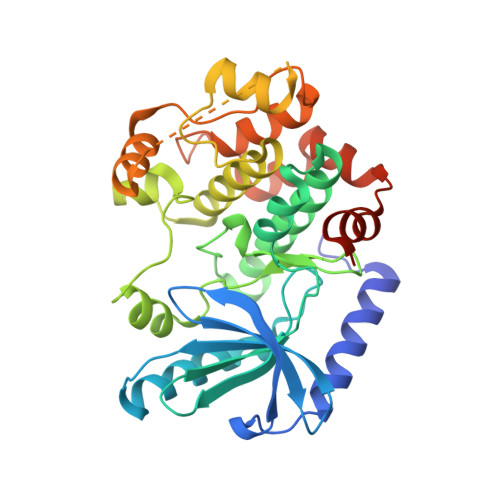
    MEK1 is a member of the MAPK signal transduction pathway that responds to growth factors and cytokines. We have determined that the kinase domain spans residues 35-382 by proteolytic cleavage. The complete kinase domain has been crystallized and its X-ray crystal structure as a complex with magnesium and ATP-gammaS determined at 2.1 A. Unlike crystals of a truncated kinase domain previously published, the crystals of the intact domain can be grown either as a binary complex with a nucleotide or as a ternary complex with a nucleotide and one of a multitude of allosteric inhibitors. Further, the crystals allow for the determination of costructures with ATP competitive inhibitors. We describe the structures of nonphosphorylated MEK1 (npMEK1) binary complexes with ADP and K252a, an ATP-competitive inhibitor (see Table 1), at 1.9 and 2.7 A resolution, respectively. Ternary complexes have also been solved between npMEK1, a nucleotide, and an allosteric non-ATP competitive inhibitor: ATP-gammaS with compound 1 and ADP with either U0126 or the MEK1 clinical candidate PD325089 at 1.8, 2.0, and 2.5 A, respectively. Compound 1 is structurally similar to PD325901. These structures illustrate fundamental differences among various mechanisms of inhibition at the molecular level. Residues 44-51 have previously been shown to play a negative regulatory role in MEK1 activity. The crystal structure of the integral kinase domain provides a structural rationale for the role of these residues. They form helix A and repress enzymatic activity by stabilizing an inactive conformation in which helix C is displaced from its active state position. Finally, the structure provides for the first time a molecular rationale that explains how mutations in MEK may lead to the cardio-facio-cutaneous syndrome.


  • Organizational Affiliation

    Schering-Plough Research Institute, Kenilworth, New Jersey 07033, USA. thierry.fischmann@spcorp.com


Macromolecules
Find similar proteins by:  (by identity cutoff)  |  3D Structure
Entity ID: 1
MoleculeChains Sequence LengthOrganismDetailsImage
Dual specificity mitogen-activated protein kinase kinase 1360Homo sapiensMutation(s): 3 
Gene Names: MAP2K1MEK1PRKMK1
EC: 2.7.12.2
UniProt & NIH Common Fund Data Resources
Find proteins for Q02750 (Homo sapiens)
Explore Q02750 
Go to UniProtKB:  Q02750
PHAROS:  Q02750
GTEx:  ENSG00000169032 
Entity Groups  
Sequence Clusters30% Identity50% Identity70% Identity90% Identity95% Identity100% Identity
UniProt GroupQ02750
Sequence Annotations
Expand
  • Reference Sequence
Small Molecules
Ligands 5 Unique
IDChains Name / Formula / InChI Key2D Diagram3D Interactions
ADP
Query on ADP

Download Ideal Coordinates CCD File 
C [auth A]ADENOSINE-5'-DIPHOSPHATE
C10 H15 N5 O10 P2
XTWYTFMLZFPYCI-KQYNXXCUSA-N
5BM
Query on 5BM

Download Ideal Coordinates CCD File 
B [auth A](2Z)-bis{amino[(2-aminophenyl)sulfanyl]methylidene}butanedinitrile
C18 H16 N6 S2
DVEXZJFMOKTQEZ-JYFOCSDGSA-N
CA
Query on CA

Download Ideal Coordinates CCD File 
E [auth A]CALCIUM ION
Ca
BHPQYMZQTOCNFJ-UHFFFAOYSA-N
MG
Query on MG

Download Ideal Coordinates CCD File 
D [auth A]MAGNESIUM ION
Mg
JLVVSXFLKOJNIY-UHFFFAOYSA-N
NA
Query on NA

Download Ideal Coordinates CCD File 
F [auth A]SODIUM ION
Na
FKNQFGJONOIPTF-UHFFFAOYSA-N
Experimental Data & Validation

Experimental Data

  • Method: X-RAY DIFFRACTION
  • Resolution: 2.00 Å
  • R-Value Work: 0.188 
  • R-Value Observed: 0.267 
  • Space Group: P 61 2 2
Unit Cell:
Length ( Å )Angle ( ˚ )
a = 76.61α = 90
b = 76.61β = 90
c = 222.9γ = 120
Software Package:
Software NamePurpose
DENZOdata reduction
SCALEPACKdata scaling
TNTrefinement
PDB_EXTRACTdata extraction

Structure Validation

View Full Validation Report


Entry History 

Deposition Data

Revision History  (Full details and data files)

  • Version 1.0: 2009-02-24
    Type: Initial release
  • Version 1.1: 2011-07-13
    Changes: Derived calculations, Version format compliance
  • Version 1.2: 2021-10-20
    Changes: Database references, Derived calculations
  • Version 1.3: 2024-02-21
    Changes: Data collection