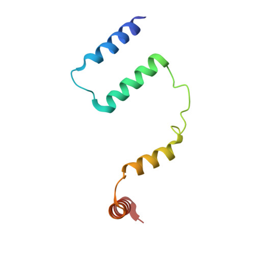
Crystal Structure of human acyl-CoA binding domain 7 complexed with palmitoyl-Coa
Kavanagh, K.L., Salah, E., Yue, W.W., Savitsky, P., Murray, J.W., Arrowsmith, C.H., Weigelt, J., Edwards, A.M., Bountra, C., von Delft, F., Oppermann, U.To be published.