3EPX

Crystal structure of Trypanosoma vivax nucleoside hydrolase in complex with the inhibitor (2R,3R,4S)-2-(hydroxymethyl)-1-(quinolin-8-ylmethyl)pyrrolidin-3,4-diol


Experimental Data Snapshot

  • Method: X-RAY DIFFRACTION
  • Resolution: 1.85 Å
  • R-Value Free: 0.210 
  • R-Value Work: 0.160 
  • R-Value Observed: 0.160 

wwPDB Validation   3D Report Full Report


Ligand Structure Quality Assessment 


This is version 1.2 of the entry. See complete history


Literature

Crystal structures of T. vivax nucleoside hydrolase in complex with new potent and specific inhibitors.

Versees, W.Goeminne, A.Berg, M.Vandemeulebroucke, A.Haemers, A.Augustyns, K.Steyaert, J.

(2009) Biochim Biophys Acta 1794: 953-960

  • DOI: https://doi.org/10.1016/j.bbapap.2009.02.011
  • Primary Citation of Related Structures:  
    3EPW, 3EPX

  • PubMed Abstract: 

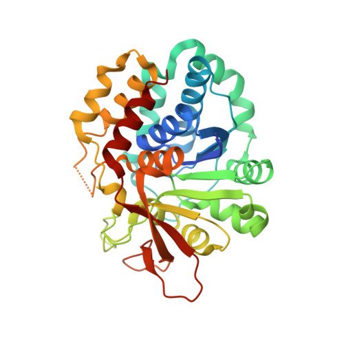
    Diseases caused by parasitic protozoa remain a major health problem, mainly due to old toxic drugs and rising drug resistance. Nucleoside hydrolases are key enzymes of the purine salvage pathway of parasites from the Trypanosomatidae family and are considered as possible drug targets. N-Arylmethyl substituted iminoribitols have been developed as selective nanomolar affinity inhibitors against the purine-specific nucleoside hydrolase of Trypanosoma vivax. The current paper describes the crystal structures of the T. vivax nucleoside hydrolase in complex with two of these inhibitors, to 1.3 and 1.85 A resolution. These high resolution structures provide an accurate picture of the mode of binding of these inhibitors and their mechanism of transition-state mimicry, and are valuable tools to guide further inhibitor design. Comparison of the current structures with previously solved structures of the enzyme in complex with ground-state and transition-state-analogue inhibitors also allows for the elucidation of a detailed molecular mechanism of active-site loop opening/closing. These loop movements can be coupled to the complex kinetic mechanism of the T. vivax nucleoside hydrolase.


  • Organizational Affiliation

    Structural Biology Brussels, Vrije Universiteit Brussel, Pleinlaan 2, B-1050 Brussels, Belgium. wversees@vub.ac.be


Macromolecules
Find similar proteins by:  (by identity cutoff)  |  3D Structure
Entity ID: 1
MoleculeChains Sequence LengthOrganismDetailsImage
IAG-nucleoside hydrolase
A, B
338Trypanosoma vivaxMutation(s): 0 
EC: 3.2.2.1
UniProt
Find proteins for Q9GPQ4 (Trypanosoma vivax)
Explore Q9GPQ4 
Go to UniProtKB:  Q9GPQ4
Entity Groups  
Sequence Clusters30% Identity50% Identity70% Identity90% Identity95% Identity100% Identity
UniProt GroupQ9GPQ4
Sequence Annotations
Expand
  • Reference Sequence
Small Molecules
Experimental Data & Validation

Experimental Data

  • Method: X-RAY DIFFRACTION
  • Resolution: 1.85 Å
  • R-Value Free: 0.210 
  • R-Value Work: 0.160 
  • R-Value Observed: 0.160 
  • Space Group: P 1 21 1
Unit Cell:
Length ( Å )Angle ( ˚ )
a = 54.422α = 90
b = 74.198β = 98.36
c = 73.553γ = 90
Software Package:
Software NamePurpose
ADSCdata collection
PHASERphasing
REFMACrefinement
DENZOdata reduction
SCALEPACKdata scaling

Structure Validation

View Full Validation Report


Ligand Structure Quality Assessment 


Entry History 

Deposition Data

Revision History  (Full details and data files)

  • Version 1.0: 2009-03-24
    Type: Initial release
  • Version 1.1: 2011-07-13
    Changes: Non-polymer description, Version format compliance
  • Version 1.2: 2023-09-06
    Changes: Data collection, Database references, Refinement description