3EOX | pdb_00003eox

High quality structure of cleaved PAI-1-stab


Experimental Data Snapshot

  • Method: X-RAY DIFFRACTION
  • Resolution: 2.61 Å
  • R-Value Free: 
    0.256 (Depositor), 0.280 (DCC) 
  • R-Value Work: 
    0.201 (Depositor) 
  • R-Value Observed: 
    0.204 (Depositor) 

Starting Model: experimental
View more details

wwPDB Validation 3D Report Full Report

Validation slider image for 3EOX

This is version 1.4 of the entry. See complete history

Literature

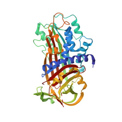
High quality structure of cleaved PAI-1-stab.

Dewilde, M.Strelkov, S.V.Rabijns, A.Declerck, P.J.

(2009) J Struct Biol 165: 126-132

  • DOI: https://doi.org/10.1016/j.jsb.2008.11.001
  • Primary Citation Related Structures: 
    3EOX

  • PubMed Abstract: 

    Here we report the crystal structure of a stablilized plasminogen activator inhibitor-1 variant (PAI-1-N150H-K154T-Q301P-Q319L-M354I (PAI-1-stab)) that shows a cleavage within the reactive centre loop. The new structure is of superior quality compared to the previously determined structure of the cleaved PAI-1-A335P mutant. We present a detailed comparison of the two structures and also compare them with the structure of the active PAI-1-stab. The structural data give important insights into the working mechanism of PAI-1 and also explain the role of various stabilizing mutations.


  • Organizational Affiliation
    • Katholieke Universiteit Leuven, Belgium.

Macromolecule Content 

  • Total Structure Weight: 42.75 kDa 
  • Atom Count: 3,084 
  • Modeled Residue Count: 370 
  • Deposited Residue Count: 379 
  • Unique protein chains: 1

Macromolecules

Find similar proteins by:|  3D Structure
Entity ID: 1
MoleculeChains  Sequence LengthOrganismDetailsImage
Plasminogen activator inhibitor 1379Homo sapiensMutation(s): 5 
Gene Names: SERPINE1PAI1PLANH1
UniProt & NIH Common Fund Data Resources
Find proteins for P05121 (Homo sapiens)
Explore P05121 
Go to UniProtKB:  P05121
PHAROS:  P05121
GTEx:  ENSG00000106366 
Entity Groups
Sequence Clusters30% Identity50% Identity70% Identity90% Identity95% Identity100% Identity
UniProt GroupP05121
Sequence Annotations
Expand
Reference Sequence

Experimental Data & Validation

Experimental Data

  • Method: X-RAY DIFFRACTION
  • Resolution: 2.61 Å
  • R-Value Free:  0.256 (Depositor), 0.280 (DCC) 
  • R-Value Work:  0.201 (Depositor) 
  • R-Value Observed: 0.204 (Depositor) 
Space Group: P 31 1 2
Unit Cell:
Length ( Å )Angle ( ˚ )
a = 71.174α = 90
b = 71.174β = 90
c = 141.664γ = 120
Software Package:
Software NamePurpose
DENZOdata reduction
SCALEPACKdata scaling
MOLREPphasing
REFMACrefinement
PDB_EXTRACTdata extraction
detectordata collection

Structure Validation

View Full Validation Report


Entry History 

Deposition Data

Revision History  (Full details and data files)

  • Version 1.0: 2009-02-17
    Type: Initial release
  • Version 1.1: 2011-07-13
    Changes: Advisory, Version format compliance
  • Version 1.2: 2017-10-25
    Changes: Advisory, Refinement description
  • Version 1.3: 2021-11-10
    Changes: Advisory, Data collection, Database references
  • Version 1.4: 2023-11-01
    Changes: Data collection, Refinement description