3EOO | pdb_00003eoo

2.9A crystal structure of methyl-isocitrate lyase from Burkholderia pseudomallei


Experimental Data Snapshot

  • Method: X-RAY DIFFRACTION
  • Resolution: 2.90 Å
  • R-Value Free: 
    0.299 (Depositor), 0.299 (DCC) 
  • R-Value Work: 
    0.237 (Depositor), 0.237 (DCC) 
  • R-Value Observed: 
    0.240 (Depositor) 

wwPDB Validation 3D Report Full Report

Validation slider image for 3EOO

This is version 1.2 of the entry. See complete history

Literature

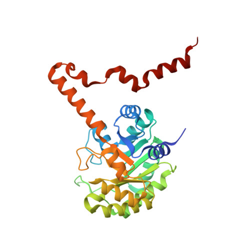
Crystal structures of isocitrate lyase from Brucella melitensis

SSGCID

To be published.

Macromolecule Content 

  • Total Structure Weight: 512.15 kDa 
  • Atom Count: 34,623 
  • Modeled Residue Count: 4,596 
  • Deposited Residue Count: 4,768 
  • Unique protein chains: 1

Macromolecules

Find similar proteins by:|  3D Structure
Entity ID: 1
MoleculeChains  Sequence LengthOrganismDetailsImage
Methylisocitrate lyase
A, B, C, D, E
A, B, C, D, E, F, G, H, I, J, K, L, M, N, O, P
298Burkholderia pseudomallei 1655Mutation(s): 0 
Gene Names: BURPS1655_C0444BURPS1710B_2458BURPS1710b_3237MSRBprpB
EC: 4.1.3.30

Experimental Data & Validation

Experimental Data

  • Method: X-RAY DIFFRACTION
  • Resolution: 2.90 Å
  • R-Value Free:  0.299 (Depositor), 0.299 (DCC) 
  • R-Value Work:  0.237 (Depositor), 0.237 (DCC) 
  • R-Value Observed: 0.240 (Depositor) 
Space Group: P 21 21 21
Unit Cell:
Length ( Å )Angle ( ˚ )
a = 163.807α = 90
b = 172.404β = 90
c = 179.277γ = 90
Software Package:
Software NamePurpose
d*TREKdata scaling
PHASERphasing
REFMACrefinement
PDB_EXTRACTdata extraction

Structure Validation

View Full Validation Report


Entry History 

Deposition Data

Revision History  (Full details and data files)

  • Version 1.0: 2008-10-21
    Type: Initial release
  • Version 1.1: 2011-07-13
    Changes: Version format compliance
  • Version 1.2: 2024-11-06
    Changes: Data collection, Database references, Refinement description, Structure summary