3EOH

Refined group II intron structure


Experimental Data Snapshot

  • Method: X-RAY DIFFRACTION
  • Resolution: 3.13 Å
  • R-Value Free: 0.237 
  • R-Value Work: 0.208 
  • R-Value Observed: 0.209 

wwPDB Validation   3D Report Full Report


This is version 1.2 of the entry. See complete history


Literature

Structural basis for exon recognition by a group II intron.

Toor, N.Rajashankar, K.Keating, K.S.Pyle, A.M.

(2008) Nat Struct Mol Biol 15: 1221-1222

  • DOI: https://doi.org/10.1038/nsmb.1509
  • Primary Citation of Related Structures:  
    3EOG, 3EOH

  • PubMed Abstract: 

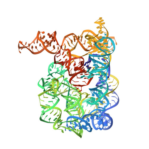
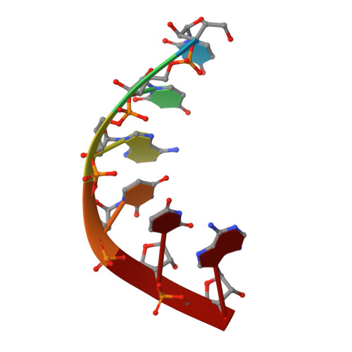
    Free group II introns are infectious retroelements that can bind and insert themselves into RNA and DNA molecules via reverse splicing. Here we report the 3.4-A crystal structure of a complex between an oligonucleotide target substrate and a group IIC intron, as well as the refined free intron structure. The structure of the complex reveals the conformation of motifs involved in exon recognition by group II introns.


  • Organizational Affiliation

    Department of Molecular Biophysics and Biochemistry, Yale University, 266 Whitney Avenue, New Haven, Connecticut 06520, USA.


Macromolecules
Find similar nucleic acids by:  (by identity cutoff)  |  3D Structure
Entity ID: 1
MoleculeChains LengthOrganismImage
Group IIC intron412N/A
Sequence Annotations
Expand
  • Reference Sequence

Find similar nucleic acids by:  Sequence   |   3D Structure  

Entity ID: 2
MoleculeChains LengthOrganismImage
5'-R(*UP*UP*AP*UP*UP*A)-3'6N/A
Sequence Annotations
Expand
  • Reference Sequence
Small Molecules
Ligands 2 Unique
IDChains Name / Formula / InChI Key2D Diagram3D Interactions
K
Query on K

Download Ideal Coordinates CCD File 
GA [auth A]
HA [auth A]
IA [auth A]
JA [auth A]
KA [auth A]
GA [auth A],
HA [auth A],
IA [auth A],
JA [auth A],
KA [auth A],
LA [auth A],
MA [auth A],
NA [auth A],
OA [auth A]
POTASSIUM ION
K
NPYPAHLBTDXSSS-UHFFFAOYSA-N
MG
Query on MG

Download Ideal Coordinates CCD File 
AA [auth A]
BA [auth A]
C [auth A]
CA [auth A]
D [auth A]
AA [auth A],
BA [auth A],
C [auth A],
CA [auth A],
D [auth A],
DA [auth A],
E [auth A],
EA [auth A],
F [auth A],
FA [auth A],
G [auth A],
H [auth A],
I [auth A],
J [auth A],
K [auth A],
L [auth A],
M [auth A],
N [auth A],
O [auth A],
P [auth A],
PA [auth B],
Q [auth A],
R [auth A],
S [auth A],
T [auth A],
U [auth A],
V [auth A],
W [auth A],
X [auth A],
Y [auth A],
Z [auth A]
MAGNESIUM ION
Mg
JLVVSXFLKOJNIY-UHFFFAOYSA-N
Experimental Data & Validation

Experimental Data

  • Method: X-RAY DIFFRACTION
  • Resolution: 3.13 Å
  • R-Value Free: 0.237 
  • R-Value Work: 0.208 
  • R-Value Observed: 0.209 
  • Space Group: P 21 21 21
Unit Cell:
Length ( Å )Angle ( ˚ )
a = 89.092α = 90
b = 94.969β = 90
c = 225.972γ = 90
Software Package:
Software NamePurpose
PHENIXrefinement

Structure Validation

View Full Validation Report


Entry History 

Deposition Data

Revision History  (Full details and data files)

  • Version 1.0: 2008-10-28
    Type: Initial release
  • Version 1.1: 2011-07-13
    Changes: Version format compliance
  • Version 1.2: 2024-02-21
    Changes: Data collection, Database references, Derived calculations