3EL6

Crystal Structure of the Erythromycin Dehydratase


Experimental Data Snapshot

  • Method: X-RAY DIFFRACTION
  • Resolution: 1.85 Å
  • R-Value Free: 0.227 
  • R-Value Work: 0.197 
  • R-Value Observed: 0.200 

wwPDB Validation   3D Report Full Report


This is version 1.2 of the entry. See complete history


Literature

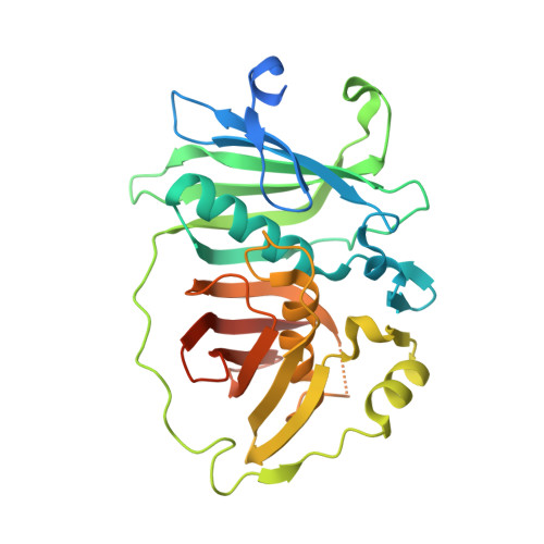
Crystal structure of the erythromycin polyketide synthase dehydratase.

Keatinge-Clay, A.

(2008) J Mol Biol 384: 941-953

  • DOI: https://doi.org/10.1016/j.jmb.2008.09.084
  • Primary Citation of Related Structures:  
    3EL6

  • PubMed Abstract: 

    The dehydratases (DHs) of modular polyketide synthases (PKSs) catalyze dehydrations that occur frequently in the biosynthesis of complex polyketides, yet little is known about them structurally or mechanistically. Here, the structure of a DH domain, isolated from the fourth module of the erythromycin PKS, is presented at 1.85 A resolution. As with the DH of the highly related animalian fatty acid synthase, the DH monomer possesses a double-hotdog fold. Two symmetry mates within the crystal lattice make a contact that likely represents the DH dimerization interface within an intact PKS. Conserved hydrophobic residues on the DH surface indicate potential interfaces with two other PKS domains, the ketoreductase and the acyl carrier protein. Mutation of an invariant arginine at the hypothesized acyl carrier protein docking site in the context of the erythromycin PKS resulted in decreased production of the erythromycin precursor 6-deoxyerythronolide B. The structure elucidates how the alpha-hydrogen and beta-hydroxyl group of a polyketide substrate interact with the catalytic histidine and aspartic acid in the DH active site prior to dehydration.


  • Organizational Affiliation

    Department of Biochemistry and Biophysics, University of California San Francisco, San Francisco, CA 94158, USA.


Macromolecules
Find similar proteins by:  (by identity cutoff)  |  3D Structure
Entity ID: 1
MoleculeChains Sequence LengthOrganismDetailsImage
Erythromycin dehydratase313Saccharopolyspora erythraeaMutation(s): 0 
Gene Names: eryA
EC: 4.2.1.61
UniProt
Find proteins for Q03132 (Saccharopolyspora erythraea)
Explore Q03132 
Go to UniProtKB:  Q03132
Entity Groups  
Sequence Clusters30% Identity50% Identity70% Identity90% Identity95% Identity100% Identity
UniProt GroupQ03132
Sequence Annotations
Expand
  • Reference Sequence
Experimental Data & Validation

Experimental Data

  • Method: X-RAY DIFFRACTION
  • Resolution: 1.85 Å
  • R-Value Free: 0.227 
  • R-Value Work: 0.197 
  • R-Value Observed: 0.200 
  • Space Group: P 43 21 2
Unit Cell:
Length ( Å )Angle ( ˚ )
a = 66.997α = 90
b = 66.997β = 90
c = 186.238γ = 90
Software Package:
Software NamePurpose
ADSCdata collection
PHENIXmodel building
REFMACrefinement
HKL-2000data reduction
HKL-2000data scaling
PHENIXphasing

Structure Validation

View Full Validation Report


Entry History 

Deposition Data

Revision History  (Full details and data files)

  • Version 1.0: 2008-11-11
    Type: Initial release
  • Version 1.1: 2011-07-13
    Changes: Version format compliance
  • Version 1.2: 2024-02-21
    Changes: Data collection, Database references, Derived calculations