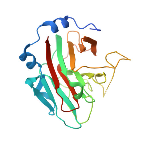
3EK9 | pdb_00003ek9

SPRY Domain-containing SOCS Box Protein 2: Crystal Structure and Residues Critical for Protein Binding


Experimental Data Snapshot

  • Method: X-RAY DIFFRACTION
  • Resolution: 2.60 Å
  • R-Value Free: 
    0.273 (Depositor), 0.270 (DCC) 
  • R-Value Work: 
    0.168 (Depositor), 0.166 (DCC) 
  • R-Value Observed: 
    0.173 (Depositor) 

Starting Model: experimental
View more details

wwPDB Validation 3D Report Full Report

Validation slider image for 3EK9

This is version 1.3 of the entry. See complete history. 

Literature

SPRY domain-containing SOCS box protein 2: crystal structure and residues critical for protein binding.

Kuang, Z., Yao, S., Xu, Y., Lewis, R.S., Low, A., Masters, S.L., Willson, T.A., Kolesnik, T.B., Nicholson, S.E., Garrett, T.J., Norton, R.S.

(2009) J Mol Biol 386: 662-674

  • DOI: https://doi.org/10.1016/j.jmb.2008.12.078
  • Primary Citation Related Structures: 
    3EK9

  • PubMed Abstract: 

    The four mammalian SPRY (a sequence repeat in dual-specificity kinase splA and ryanodine receptors) domain-containing suppressor of cytokine signalling (SOCS) box proteins (SSB-1 to -4) are characterised by a C-terminal SOCS box and a central SPRY domain. The latter is a protein interaction module found in over 1600 proteins, with more than 70 encoded in the human genome. Here we report the crystal structure of the SPRY domain of murine SSB-2 and compare it with the SSB-2 solution structure and crystal structures of other B30.2/SPRY domain-containing family proteins. The structure is a bent beta-sandwich, consisting of two seven-stranded beta-sheets wrapped around a long loop that extends from the centre strands of the inner or concave beta-sheet; it closely matches those of GUSTAVUS and SSB-4. The structure is also similar to those of two recently determined Neuralized homology repeat (NHR) domains (also known as NEUZ domains), with detailed comparisons, suggesting that the NEUZ/NHR domains form a subclass of SPRY domains. The binding site on SSB-2 for the prostate apoptosis response-4 (Par-4) protein has been mapped in finer detail using mutational analyses. Moreover, SSB-1 was shown to have a Par-4 binding surface similar to that identified for SSB-2. Structural perturbations of SSB-2 induced by mutations affecting its interaction with Par-4 and/or c-Met have been characterised by NMR. These comparisons, in conjunction with previously published dynamics data from NMR relaxation studies and coarse-grained dynamics simulation using normal mode analysis, further refine our understanding of the structural basis for protein recognition of SPRY domain-containing proteins.


  • Organizational Affiliation: 
    • The Walter and Eliza Hall Institute of Medical Research, 1G Royal Parade, Parkville, Victoria 3052, Australia.

Macromolecule Content 

  • Total Structure Weight: 23.46 kDa 
  • Atom Count: 1,541 
  • Modeled Residue Count: 193 
  • Deposited Residue Count: 213 
  • Unique protein chains: 1

Macromolecules

Find similar proteins by:|  3D Structure
Entity ID: 1
MoleculeChains  Sequence LengthOrganismDetailsImage
SPRY domain-containing SOCS box protein 2213Mus musculusMutation(s): 0 
Gene Names: Spsb2, Grcc9, Ssb2
UniProt
Find proteins for O88838 (Mus musculus)
Explore O88838 
Go to UniProtKB:  O88838
Entity Groups
Sequence Clusters30% Identity50% Identity70% Identity90% Identity95% Identity100% Identity
UniProt GroupO88838
Sequence Annotations
Expand
Reference Sequence

Small Molecules

Ligands 1 Unique
IDChains Name / Formula / InChI Key2D Diagram3D Interactions
GOL

Query on GOL


Download:Ideal Coordinates CCD File
B [auth A]GLYCEROL
C3 H8 O3
PEDCQBHIVMGVHV-UHFFFAOYSA-N

Experimental Data & Validation

Experimental Data

  • Method: X-RAY DIFFRACTION
  • Resolution: 2.60 Å
  • R-Value Free:  0.273 (Depositor), 0.270 (DCC) 
  • R-Value Work:  0.168 (Depositor), 0.166 (DCC) 
  • R-Value Observed: 0.173 (Depositor) 
Space Group: P 21 21 21
Unit Cell:
Length ( Å )Angle ( ˚ )
a = 34.255α = 90
b = 68.176β = 90
c = 69.012γ = 90
Software Package:
Software NamePurpose
CrystalCleardata collection
MOLREPphasing
REFMACrefinement
HKL-2000data reduction
HKL-2000data scaling

Structure Validation

View Full Validation Report


Entry History 

Deposition Data

Revision History  (Full details and data files)

  • Version 1.0: 2009-02-24
    Type: Initial release
  • Version 1.1: 2011-07-13
    Changes: Non-polymer description, Version format compliance
  • Version 1.2: 2012-02-08
    Changes: Structure summary
  • Version 1.3: 2023-08-30
    Changes: Data collection, Database references, Derived calculations, Refinement description