3EJG

Crystal structure of HCoV-229E X-domain


Experimental Data Snapshot

  • Method: X-RAY DIFFRACTION
  • Resolution: 1.78 Å
  • R-Value Free: 0.205 
  • R-Value Work: 0.165 
  • R-Value Observed: 0.167 

wwPDB Validation   3D Report Full Report


This is version 1.3 of the entry. See complete history


Literature

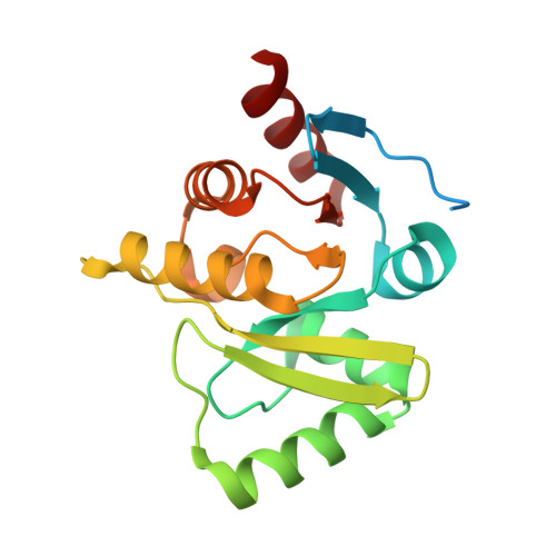
Crystal structures of the X-domains of a Group-1 and a Group-3 coronavirus reveal that ADP-ribose-binding may not be a conserved property.

Piotrowski, Y.Hansen, G.Boomaars-van der Zanden, A.L.Snijder, E.J.Gorbalenya, A.E.Hilgenfeld, R.

(2009) Protein Sci 18: 6-16

  • DOI: https://doi.org/10.1002/pro.15
  • Primary Citation of Related Structures:  
    3EJF, 3EJG, 3EKE

  • PubMed Abstract: 

    The polyproteins of coronaviruses are cleaved by viral proteases into at least 15 nonstructural proteins (Nsps). Consisting of five domains, Nsp3 is the largest of these (180-210 kDa). Among these domains, the so-called X-domain is believed to act as ADP-ribose-1''-phosphate phosphatase or to bind poly(ADP-ribose). However, here we show that the X-domain of Infectious Bronchitis Virus (strain Beaudette), a Group-3 coronavirus, fails to bind ADP-ribose. This is explained on the basis of the crystal structure of the protein, determined at two different pH values. For comparison, we also describe the crystal structure of the homologous X-domain from Human Coronavirus 229E, a Group-1 coronavirus, which does bind ADP-ribose.


  • Organizational Affiliation

    Institute of Biochemistry, Center for Structural and Cell Biology in Medicine, University of Lübeck, Ratzeburger Allee 160, 23538 Lübeck, Germany.


Macromolecules
Find similar proteins by:  (by identity cutoff)  |  3D Structure
Entity ID: 1
MoleculeChains Sequence LengthOrganismDetailsImage
Non-structural protein 3193Human coronavirus 229EMutation(s): 0 
Gene Names: 1a
EC: 3.4.22
UniProt
Find proteins for P0C6X1 (Human coronavirus 229E)
Explore P0C6X1 
Go to UniProtKB:  P0C6X1
Entity Groups  
Sequence Clusters30% Identity50% Identity70% Identity90% Identity95% Identity100% Identity
UniProt GroupP0C6X1
Sequence Annotations
Expand
  • Reference Sequence
Experimental Data & Validation

Experimental Data

  • Method: X-RAY DIFFRACTION
  • Resolution: 1.78 Å
  • R-Value Free: 0.205 
  • R-Value Work: 0.165 
  • R-Value Observed: 0.167 
  • Space Group: P 1 21 1
Unit Cell:
Length ( Å )Angle ( ˚ )
a = 33.56α = 90
b = 65.89β = 110.07
c = 38.02γ = 90
Software Package:
Software NamePurpose
SCALAdata scaling
REFMACrefinement
PDB_EXTRACTdata extraction
MOSFLMdata reduction
PHASERphasing

Structure Validation

View Full Validation Report


Entry History 

Deposition Data

Revision History  (Full details and data files)

  • Version 1.0: 2008-09-30
    Type: Initial release
  • Version 1.1: 2011-07-13
    Changes: Version format compliance
  • Version 1.2: 2018-03-07
    Changes: Data collection
  • Version 1.3: 2024-02-21
    Changes: Data collection, Database references