


Human dUTPase in complex with alpha,beta-imido-dUTP and Mg2+: visualization of the full-length C-termini in all monomers and suggestion for an additional metal ion binding site
Takacs, E., Barabas, O., Vertessy, B.G.To be published.
Experimental Data Snapshot
Starting Model: experimental
View more details
Entity ID: 1 | |||||
|---|---|---|---|---|---|
| Molecule | Chains | Sequence Length | Organism | Details | Image |
| dUTP pyrophosphatase | 164 | Homo sapiens | Mutation(s): 0 Gene Names: DUT EC: 3.6.1.23 | 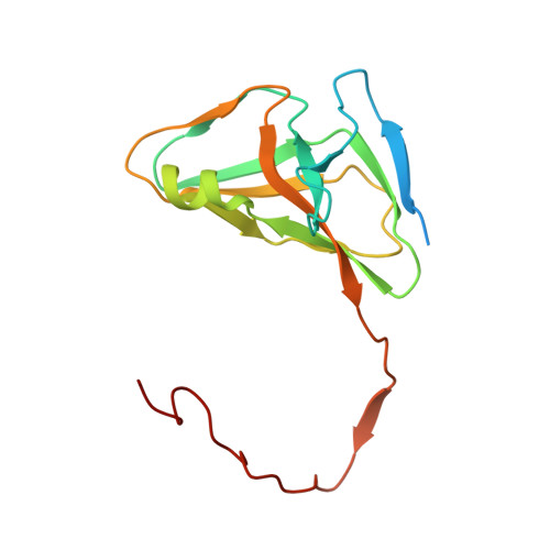 | |
UniProt & NIH Common Fund Data Resources | |||||
PHAROS: P33316 GTEx: ENSG00000128951 | |||||
Entity Groups | |||||
| Sequence Clusters | 30% Identity50% Identity70% Identity90% Identity95% Identity100% Identity | ||||
| UniProt Group | P33316 | ||||
Sequence AnnotationsExpand | |||||
Reference Sequence | |||||
| Ligands 2 Unique | |||||
|---|---|---|---|---|---|
| ID | Chains | Name / Formula / InChI Key | 2D Diagram | 3D Interactions | |
| DUP Download:Ideal Coordinates CCD File | H [auth A] K [auth B] L [auth C] O [auth X] R [auth Y] | 2'-DEOXYURIDINE 5'-ALPHA,BETA-IMIDO-TRIPHOSPHATE C9 H16 N3 O13 P3 XZLLMTSKYYYJLH-SHYZEUOFSA-N |  | ||
| MG Download:Ideal Coordinates CCD File | G [auth A] I [auth A] J [auth B] M [auth C] N [auth X] | MAGNESIUM ION Mg JLVVSXFLKOJNIY-UHFFFAOYSA-N |  | ||
| Length ( Å ) | Angle ( ˚ ) |
|---|---|
| a = 65.446 | α = 90 |
| b = 87.16 | β = 90.09 |
| c = 70.608 | γ = 90 |
| Software Name | Purpose |
|---|---|
| MOLREP | phasing |
| REFMAC | refinement |
| XDS | data reduction |
| XSCALE | data scaling |


