


Crystal structure of Pru du amandin, an allergenic protein from prunus dulcis
Gaur, V., Salunke, D.M.To be published.
Experimental Data Snapshot
Starting Model: experimental
View more details
wwPDB Validation 3D Report Full Report

Entity ID: 1 | |||||
|---|---|---|---|---|---|
| Molecule | Chains | Sequence Length | Organism | Details | Image |
| Prunin | 531 | Prunus dulcis | Mutation(s): 0 | 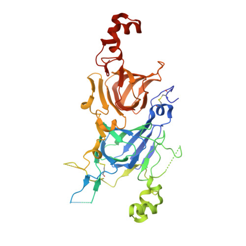 | |
UniProt | |||||
Entity Groups | |||||
| Sequence Clusters | 30% Identity50% Identity70% Identity90% Identity95% Identity100% Identity | ||||
| UniProt Group | Q43607 | ||||
Sequence AnnotationsExpand | |||||
Reference Sequence | |||||
| Length ( Å ) | Angle ( ˚ ) |
|---|---|
| a = 151.099 | α = 90 |
| b = 151.099 | β = 90 |
| c = 164.628 | γ = 90 |
| Software Name | Purpose |
|---|---|
| MAR345dtb | data collection |
| AMoRE | phasing |
| REFMAC | refinement |
| AUTOMAR | data reduction |


