3EGA

Crystal structure of Pellino2 FHA Domain at 1.8 Angstroms resolution


Experimental Data Snapshot

  • Method: X-RAY DIFFRACTION
  • Resolution: 1.80 Å
  • R-Value Free: 0.244 
  • R-Value Work: 0.210 
  • R-Value Observed: 0.212 

wwPDB Validation   3D Report Full Report


This is version 1.4 of the entry. See complete history


Literature

Pellino proteins contain a cryptic FHA domain that mediates interaction with phosphorylated IRAK1.

Lin, C.C.Huoh, Y.S.Schmitz, K.R.Jensen, L.E.Ferguson, K.M.

(2008) Structure 16: 1806-1816

  • DOI: https://doi.org/10.1016/j.str.2008.09.011
  • Primary Citation of Related Structures:  
    3EGA, 3EGB

  • PubMed Abstract: 

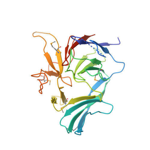
    Pellino proteins are RING E3 ubiquitin ligases involved in signaling events downstream of the Toll and interleukin-1 (IL-1) receptors, key initiators of innate immune and inflammatory responses. Pellino proteins associate with and ubiquitinate proteins in these pathways, including the interleukin-1 receptor associated kinase-1 (IRAK1). We determined the X-ray crystal structure of a Pellino2 fragment lacking only the RING domain. This structure reveals that the IRAK1-binding region of Pellino proteins consists largely of a previously unidentified forkhead-associated (FHA) domain. FHA domains are well-characterized phosphothreonine-binding modules, and this cryptic example in Pellino2 can drive interaction of this protein with phosphorylated IRAK1. The Pellino FHA domain is decorated with an unusual appendage or "wing" composed of two long inserts that lie within the FHA homology region. Delineating how this E3 ligase associates with substrates, and how these interactions are regulated by phosphorylation, is crucial for a complete understanding of Toll/IL-1 receptor signaling.


  • Organizational Affiliation

    Department of Physiology, University of Pennsylvania School of Medicine, Philadelphia, PA 19104, USA.


Macromolecules
Find similar proteins by:  (by identity cutoff)  |  3D Structure
Entity ID: 1
MoleculeChains Sequence LengthOrganismDetailsImage
Protein pellino homolog 2263Homo sapiensMutation(s): 2 
Gene Names: PELI2
UniProt & NIH Common Fund Data Resources
Find proteins for Q9HAT8 (Homo sapiens)
Explore Q9HAT8 
Go to UniProtKB:  Q9HAT8
PHAROS:  Q9HAT8
GTEx:  ENSG00000139946 
Entity Groups  
Sequence Clusters30% Identity50% Identity70% Identity90% Identity95% Identity100% Identity
UniProt GroupQ9HAT8
Sequence Annotations
Expand
  • Reference Sequence
Small Molecules
Ligands 1 Unique
IDChains Name / Formula / InChI Key2D Diagram3D Interactions
SO4
Query on SO4

Download Ideal Coordinates CCD File 
B [auth A]SULFATE ION
O4 S
QAOWNCQODCNURD-UHFFFAOYSA-L
Modified Residues  1 Unique
IDChains TypeFormula2D DiagramParent
MSE
Query on MSE
A
L-PEPTIDE LINKINGC5 H11 N O2 SeMET
Experimental Data & Validation

Experimental Data

  • Method: X-RAY DIFFRACTION
  • Resolution: 1.80 Å
  • R-Value Free: 0.244 
  • R-Value Work: 0.210 
  • R-Value Observed: 0.212 
  • Space Group: F 2 2 2
Unit Cell:
Length ( Å )Angle ( ˚ )
a = 83.65α = 90
b = 86.137β = 90
c = 162.767γ = 90
Software Package:
Software NamePurpose
DENZOdata reduction
SCALEPACKdata scaling
SHELXphasing
REFMACrefinement
PDB_EXTRACTdata extraction
ADSCdata collection
HKL-3000data reduction
HKL-3000data scaling
SHELXDphasing

Structure Validation

View Full Validation Report


Entry History 

Deposition Data

Revision History  (Full details and data files)

  • Version 1.0: 2008-12-23
    Type: Initial release
  • Version 1.1: 2011-07-13
    Changes: Version format compliance
  • Version 1.2: 2012-03-21
    Changes: Database references
  • Version 1.3: 2017-10-25
    Changes: Refinement description
  • Version 1.4: 2021-10-20
    Changes: Database references, Derived calculations