3EDC | pdb_00003edc

Crystal Structure of a 1.6-hexanediol Bound Tetrameric Form of Escherichia coli Lac-repressor Refined to 2.1 Resolution


Experimental Data Snapshot

  • Method: X-RAY DIFFRACTION
  • Resolution: 2.10 Å
  • R-Value Free: 
    0.201 (Depositor), 0.219 (DCC) 
  • R-Value Work: 
    0.156 (Depositor) 
  • R-Value Observed: 
    0.159 (Depositor) 

Starting Model: experimental
View more details

wwPDB Validation 3D Report Full Report

Validation slider image for 3EDC

This is version 1.2 of the entry. See complete history

Literature

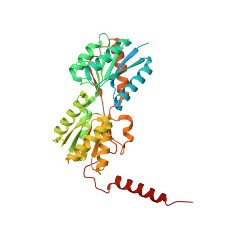
Crystal structure of a 1.6-hexanediol bound tetrameric form of Escherichia coli lac-repressor refined to 2.1 A resolution

Stenberg, K.A.E.Vihinen, M.

(2009) Proteins 75: 748-759

  • DOI: https://doi.org/10.1002/prot.22284
  • Primary Citation Related Structures: 
    3EDC

  • PubMed Abstract: 

    We report the structure of a novel tetrameric form of the lactose repressor (LacI) protein from Escherichia coli refined to 2.1 A resolution. The tetramer is bound to 1.6-hexanediol present in the crystallization solution and the final R(free) for the structure is 0.201. The structure confirms previously reported structures on the monomer level. However, the tetramer is much more densely packed. This adds a new level of complexity to the interpretation of mutational effects and challenges details in the current model for LacI function. Several amino acids, previously associated with changes in function but unexplained at the structural level, appear in a new structural context in this tetramer which provides new implications for their function.


  • Organizational Affiliation
    • Faculty of Biosciences, Division of Biochemistry, University of Helsinki, Helsinki, Finland. kaj.stenberg@helsinki.fi

Macromolecule Content 

  • Total Structure Weight: 154.98 kDa 
  • Atom Count: 9,549 
  • Modeled Residue Count: 1,200 
  • Deposited Residue Count: 1,440 
  • Unique protein chains: 1

Macromolecules

Find similar proteins by:|  3D Structure
Entity ID: 1
MoleculeChains  Sequence LengthOrganismDetailsImage
Lactose operon repressor
A, B, C, D
360Escherichia coli K-12Mutation(s): 0 
UniProt
Find proteins for P03023 (Escherichia coli (strain K12))
Explore P03023 
Go to UniProtKB:  P03023
Entity Groups
Sequence Clusters30% Identity50% Identity70% Identity90% Identity95% Identity100% Identity
UniProt GroupP03023
Sequence Annotations
Expand
Reference Sequence

Experimental Data & Validation

Experimental Data

  • Method: X-RAY DIFFRACTION
  • Resolution: 2.10 Å
  • R-Value Free:  0.201 (Depositor), 0.219 (DCC) 
  • R-Value Work:  0.156 (Depositor) 
  • R-Value Observed: 0.159 (Depositor) 
Space Group: C 1 2 1
Unit Cell:
Length ( Å )Angle ( ˚ )
a = 164.15α = 90
b = 72.434β = 113.96
c = 130.566γ = 90
Software Package:
Software NamePurpose
HKL-2000data collection
PHASESphasing
REFMACrefinement
DENZOdata reduction
SCALEPACKdata scaling

Structure Validation

View Full Validation Report


Entry History 

Deposition Data

Revision History  (Full details and data files)

  • Version 1.0: 2008-11-25
    Type: Initial release
  • Version 1.1: 2011-07-13
    Changes: Advisory, Version format compliance
  • Version 1.2: 2023-11-01
    Changes: Data collection, Database references, Derived calculations, Refinement description