3ECY | pdb_00003ecy

Crystal structural analysis of Drosophila melanogaster dUTPase


Experimental Data Snapshot

  • Method: X-RAY DIFFRACTION
  • Resolution: 1.88 Å
  • R-Value Free: 
    0.222 (Depositor), 0.226 (DCC) 
  • R-Value Work: 
    0.200 (Depositor), 0.206 (DCC) 
  • R-Value Observed: 
    0.202 (Depositor) 

Starting Model: experimental
View more details

wwPDB Validation 3D Report Full Report

Validation slider image for 3ECY

This is version 1.2 of the entry. See complete history

Literature

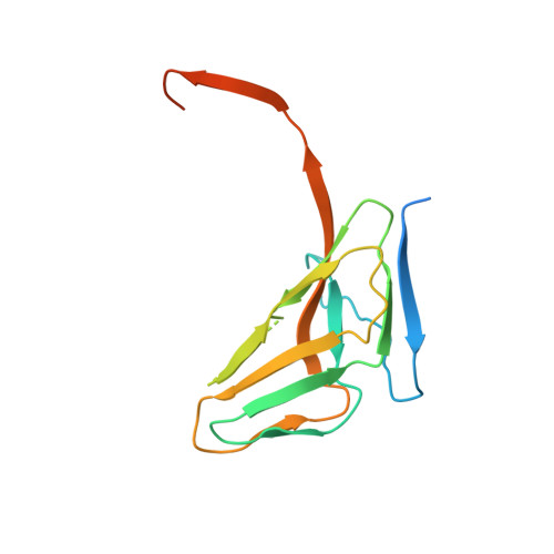
Molecular shape and prominent role of beta-strand swapping in organization of dUTPase oligomers.

Takacs, E.Barabas, O.Petoukhov, M.V.Svergun, D.I.Vertessy, B.G.

(2009) FEBS Lett 583: 865-871

  • DOI: https://doi.org/10.1016/j.febslet.2009.02.011
  • Primary Citation Related Structures: 
    3ECY

  • PubMed Abstract: 

    Most dUTP pyrophosphatases (dUTPases) are homotrimers with interfaces formed between subunit surfaces, in the central channel, and by C-terminal beta-strand swapping. Analysis of intersubunit interactions reveals an important cohesive role for the C-terminus. This is reflected in the crystal structure of fruitfly dUTPase displaying a dimeric organization in crystals grown in alcohol solution, where only beta-strand swapping interactions between subunits are retained from the usual trimer structure. Mutations of a suggested hinge proline destabilize human and Escherichia coli dUTPases without preventing trimeric organization. Trimer formation was, however, prevented in the human enzyme by truncating the C-terminus before the swapping arm. The molecular shape of full-length enzymes in solution reveals the localization and variation in flexibility of N- and C-terminal segments.


  • Organizational Affiliation
    • Institute of Enzimology, BRC, Hungarian Academy of Sciences, Budapest, Hungary.

Macromolecule Content 

  • Total Structure Weight: 34.71 kDa 
  • Atom Count: 1,907 
  • Modeled Residue Count: 238 
  • Deposited Residue Count: 320 
  • Unique protein chains: 1

Macromolecules

Find similar proteins by:|  3D Structure
Entity ID: 1
MoleculeChains  Sequence LengthOrganismDetailsImage
CG4584-PA, isoform A (BcDNA.LD08534)
A, B
160Drosophila melanogasterMutation(s): 0 
Gene Names: dUTPaseBcDNA.LD08534CG4584Dmel_CG4584
EC: 3.6.1.23
UniProt
Find proteins for Q9V3I1 (Drosophila melanogaster)
Explore Q9V3I1 
Go to UniProtKB:  Q9V3I1
Entity Groups
Sequence Clusters30% Identity50% Identity70% Identity90% Identity95% Identity100% Identity
UniProt GroupQ9V3I1
Sequence Annotations
Expand
Reference Sequence

Experimental Data & Validation

Experimental Data

  • Method: X-RAY DIFFRACTION
  • Resolution: 1.88 Å
  • R-Value Free:  0.222 (Depositor), 0.226 (DCC) 
  • R-Value Work:  0.200 (Depositor), 0.206 (DCC) 
  • R-Value Observed: 0.202 (Depositor) 
Space Group: P 41
Unit Cell:
Length ( Å )Angle ( ˚ )
a = 42.799α = 90
b = 42.799β = 90
c = 169.384γ = 90
Software Package:
Software NamePurpose
REFMACrefinement
XDSdata reduction
XSCALEdata scaling
MOLREPphasing

Structure Validation

View Full Validation Report


Entry History 

Deposition Data

Revision History  (Full details and data files)

  • Version 1.0: 2008-10-07
    Type: Initial release
  • Version 1.1: 2011-07-13
    Changes: Advisory, Version format compliance
  • Version 1.2: 2023-08-30
    Changes: Data collection, Database references, Refinement description