Structural and biochemical characterization of MepR, a multidrug binding transcription regulator of the Staphylococcus aureus multidrug efflux pump MepA.
Kumaraswami, M., Schuman, J.T., Seo, S.M., Kaatz, G.W., Brennan, R.G.(2009) Nucleic Acids Res 37: 1211-1224
- PubMed: 19129225 Search on PubMedSearch on PubMed Central
- DOI: https://doi.org/10.1093/nar/gkn1046
- Primary Citation Related Structures:
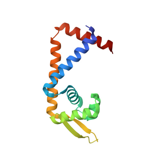
3ECO - PubMed Abstract:
MepR is a multidrug binding transcription regulator that represses expression of the Staphylococcus aureus multidrug efflux pump gene, mepA, as well as its own gene. MepR is induced by multiple cationic toxins, which are also substrates of MepA. In order to understand the gene regulatory and drug-binding mechanisms of MepR, we carried out biochemical, in vivo and structural studies. The 2.40 A resolution structure of drug-free MepR reveals the most open MarR family protein conformation to date, which will require a huge conformational change to bind cognate DNA. DNA-binding data show that MepR uses a dual regulatory binding mode as the repressor binds the mepA operator as a dimer of dimers, but binds the mepR operator as a single dimer. Alignment of the six half sites reveals the consensus MepR binding site, 5'-GTTAGAT-3'. 'Drug' binding studies show that MepR binds to ethidium and DAPI with comparable affinities (K(d) = 2.6 and 4.5 microM, respectively), but with significantly lower affinity to the larger rhodamine 6G (K(d) = 62.6 microM). Mapping clinically relevant or in vitro selected MepR mutants onto the MepR structure suggests that their defective repressor phenotypes are due to structural and allosteric defects.
- Department of Biochemistry and Molecular Biology, University of Texas M.D. Anderson Cancer Center, Houston, TX 77030, USA.
Organizational Affiliation: