3ECC | pdb_00003ecc

Crystal structure of the DnaC helicase loader in complex with ADP-BeF3


Experimental Data Snapshot

  • Method: X-RAY DIFFRACTION
  • Resolution: 2.70 Å
  • R-Value Free: 
    0.294 (Depositor), 0.300 (DCC) 
  • R-Value Work: 
    0.263 (Depositor), 0.260 (DCC) 
  • R-Value Observed: 
    0.263 (Depositor) 

wwPDB Validation 3D Report Full Report

Validation slider image for 3ECC

Ligand Structure Quality Assessment 


This is version 1.3 of the entry. See complete history

Literature

Structural synergy and molecular crosstalk between bacterial helicase loaders and replication initiators.

Mott, M.L.Erzberger, J.P.Coons, M.M.Berger, J.M.

(2008) Cell 135: 623-634

  • DOI: https://doi.org/10.1016/j.cell.2008.09.058
  • Primary Citation Related Structures: 
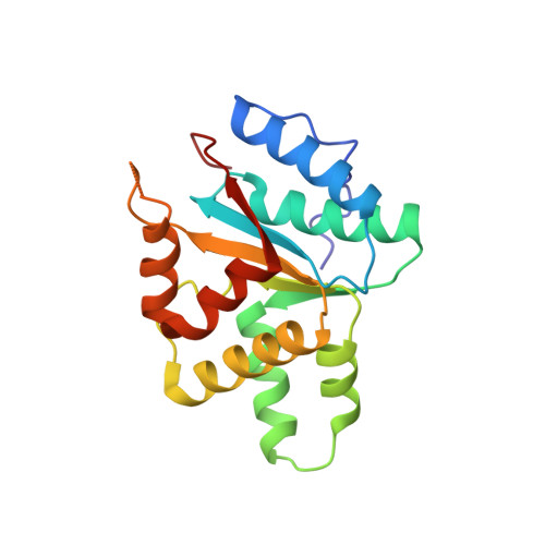
    3EC2, 3ECC

  • PubMed Abstract: 

    The loading of oligomeric helicases onto replication origins marks an essential step in replisome assembly. In cells, dedicated AAA+ ATPases regulate loading, however, the mechanism by which these factors recruit and deposit helicases has remained unclear. To better understand this process, we determined the structure of the ATPase region of the bacterial helicase loader DnaC from Aquifex aeolicus to 2.7 A resolution. The structure shows that DnaC is a close paralog of the bacterial replication initiator, DnaA, and unexpectedly shares an ability to form a helical assembly similar to that of ATP-bound DnaA. Complementation and ssDNA-binding assays validate the importance of homomeric DnaC interactions, while pull-down experiments show that the DnaC and DnaA AAA+ domains interact in a nucleotide-dependent manner. These findings implicate DnaC as a molecular adaptor that uses ATP-activated DnaA as a docking site for regulating the recruitment and correct spatial deposition of the DnaB helicase onto origins.


  • Organizational Affiliation
    • Molecular and Cell Biology Department, Quantitative Biosciences Institute, University of California, Berkeley, CA 94720, USA.

Macromolecule Content 

  • Total Structure Weight: 21.77 kDa 
  • Atom Count: 1,548 
  • Modeled Residue Count: 182 
  • Deposited Residue Count: 185 
  • Unique protein chains: 1

Macromolecules

Find similar proteins by:|  3D Structure
Entity ID: 1
MoleculeChains  Sequence LengthOrganismDetailsImage
DNA replication protein DnaC185Aquifex aeolicusMutation(s): 0 
Gene Names: dnaCaq_910
UniProt
Find proteins for O67056 (Aquifex aeolicus (strain VF5))
Explore O67056 
Go to UniProtKB:  O67056
Entity Groups
Sequence Clusters30% Identity50% Identity70% Identity90% Identity95% Identity100% Identity
UniProt GroupO67056
Sequence Annotations
Expand
Reference Sequence

Experimental Data & Validation

Experimental Data

  • Method: X-RAY DIFFRACTION
  • Resolution: 2.70 Å
  • R-Value Free:  0.294 (Depositor), 0.300 (DCC) 
  • R-Value Work:  0.263 (Depositor), 0.260 (DCC) 
  • R-Value Observed: 0.263 (Depositor) 
Space Group: P 61
Unit Cell:
Length ( Å )Angle ( ˚ )
a = 84.582α = 90
b = 84.582β = 90
c = 49.587γ = 120
Software Package:
Software NamePurpose
ELVESrefinement
REFMACrefinement
DENZOdata reduction
SCALAdata scaling
REFMACphasing

Structure Validation

View Full Validation Report


Ligand Structure Quality Assessment 


Entry History 

Deposition Data

Revision History  (Full details and data files)

  • Version 1.0: 2008-11-25
    Type: Initial release
  • Version 1.1: 2011-07-13
    Changes: Version format compliance
  • Version 1.2: 2017-10-25
    Changes: Refinement description
  • Version 1.3: 2024-02-21
    Changes: Data collection, Database references, Derived calculations