3EBX

REFINEMENT AT 1.4 ANGSTROMS RESOLUTION OF A MODEL OF ERABUTOXIN B. TREATMENT OF ORDERED SOLVENT AND DISCRETE DISORDER


Experimental Data Snapshot

  • Method: X-RAY DIFFRACTION
  • Resolution: 1.40 Å
  • R-Value Observed: 0.140 

wwPDB Validation   3D Report Full Report


This is version 1.3 of the entry. See complete history


Literature

Refinement at 1.4 A resolution of a model of erabutoxin b: treatment of ordered solvent and discrete disorder.

Smith, J.L.Corfield, P.W.Hendrickson, W.A.Low, B.W.

(1988) Acta Crystallogr A 44: 357-368

  • DOI: https://doi.org/10.1107/s0108767388000303
  • Primary Citation of Related Structures:  
    3EBX

  • PubMed Abstract: 

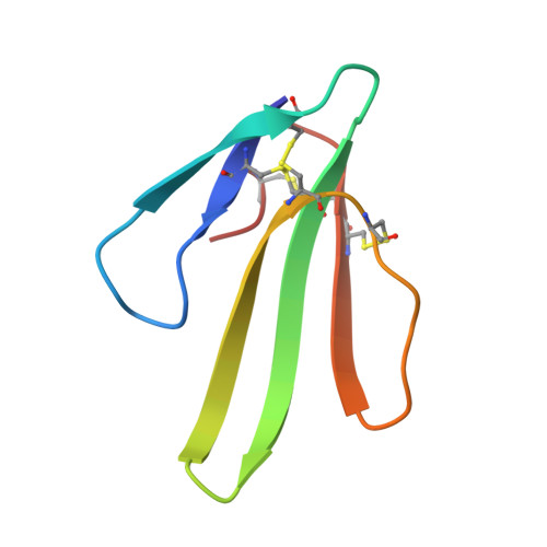
    The latter stages in the refinement of the protein erabutoxin b are described. The crystal structure of the 62-residue protein has been refined to a conventional R factor of 0.144 by stereochemically restrained least-squares methods using diffraction data to a limit of 1.4 A spacings. Emphasis was placed on determining as accurately as possible the solvent structure and the structures of heterogeneous groups in the protein. The final model includes two conformers for each of seven side chains and for an octapeptide segment. A total of 111 sites for water molecules have been located as well as one sulfate ion with a total of 68 site occupancies. 65 of the solvent sites overlap either with protein atoms belonging to groups in two alternative conformations or with other solvent sites. Dual protein conformers and overlapping solvent sites were both included in the least-squares refinement. Individual thermal and occupancy parameters were refined for solvent molecules. An analysis of these parameters has provided useful structural information.


  • Organizational Affiliation

    Department of Biochemistry and Molecular Biophysics, Columbia University, New York, NY 10032.


Macromolecules
Find similar proteins by:  (by identity cutoff)  |  3D Structure
Entity ID: 1
MoleculeChains Sequence LengthOrganismDetailsImage
ERABUTOXIN B62Laticauda semifasciataMutation(s): 0 
Membrane Entity: Yes 
UniProt
Find proteins for Q90VW1 (Laticauda semifasciata)
Explore Q90VW1 
Go to UniProtKB:  Q90VW1
Entity Groups  
Sequence Clusters30% Identity50% Identity70% Identity90% Identity95% Identity100% Identity
UniProt GroupQ90VW1
Sequence Annotations
Expand
  • Reference Sequence
Small Molecules
Ligands 1 Unique
IDChains Name / Formula / InChI Key2D Diagram3D Interactions
SO4
Query on SO4

Download Ideal Coordinates CCD File 
B [auth A]SULFATE ION
O4 S
QAOWNCQODCNURD-UHFFFAOYSA-L
Experimental Data & Validation

Experimental Data

  • Method: X-RAY DIFFRACTION
  • Resolution: 1.40 Å
  • R-Value Observed: 0.140 
  • Space Group: P 21 21 21
Unit Cell:
Length ( Å )Angle ( ˚ )
a = 49.94α = 90
b = 46.58β = 90
c = 21.59γ = 90
Software Package:
Software NamePurpose
PROLSQrefinement

Structure Validation

View Full Validation Report


Entry History 

Deposition Data

Revision History  (Full details and data files)

  • Version 1.0: 1988-04-16
    Type: Initial release
  • Version 1.1: 2008-03-03
    Changes: Version format compliance
  • Version 1.2: 2011-07-13
    Changes: Version format compliance
  • Version 1.3: 2017-11-29
    Changes: Derived calculations, Other