3EB9

Crystal structure of 6-phosphogluconolactonase from trypanosoma brucei complexed with citrate


Experimental Data Snapshot

  • Method: X-RAY DIFFRACTION
  • Resolution: 2.00 Å
  • R-Value Free: 0.244 
  • R-Value Work: 0.195 
  • R-Value Observed: 0.198 

wwPDB Validation   3D Report Full Report


This is version 1.3 of the entry. See complete history


Literature

Insights into the enzymatic mechanism of 6-phosphogluconolactonase from Trypanosoma brucei using structural data and molecular dynamics simulation.

Duclert-Savatier, N.Poggi, L.Miclet, E.Lopes, P.Ouazzani, J.Chevalier, N.Nilges, M.Delarue, M.Stoven, V.

(2009) J Mol Biol 388: 1009-1021

  • DOI: https://doi.org/10.1016/j.jmb.2009.03.063
  • Primary Citation of Related Structures:  
    3E7F, 3EB9

  • PubMed Abstract: 

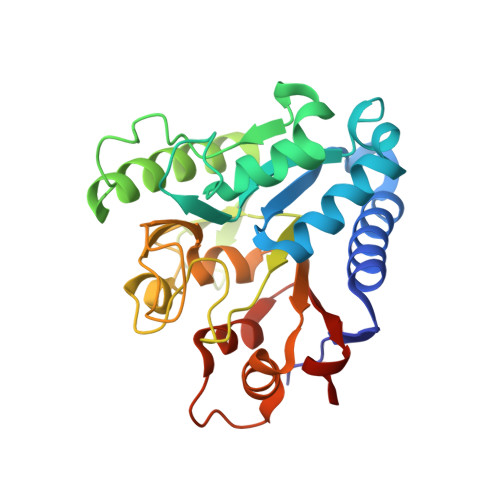
    Trypanosoma brucei is the causative agent of African sleeping sickness. Current work for the development of new drugs against this pathology includes evaluation of enzymes of the pentose phosphate pathway (PPP), which first requires a clear understanding of their function and mechanism of action. In this context, we focused on T. brucei 6-phosphogluconolactonase (Tb6PGL), which converts delta-6-phosphogluconolactone into 6-phosphogluconic acid in the second step of the PPP. We have determined the crystal structure of Tb6PGL in complex with two ligands, 6-phosphogluconic acid and citrate, at 2.2 A and 2.0 A resolution, respectively. We have performed molecular dynamics (MD) simulations on Tb6PGL in its empty form and in complex with delta-6-phosphogluconolactone, its natural ligand. Analysis of the structural data and MD simulations allowed us to propose a detailed enzymatic mechanism for 6PGL enzymes.


  • Organizational Affiliation

    Unité de Bioinformatique Structurale, Institut Pasteur, URA 2185 du CNRS, Paris, France.


Macromolecules
Find similar proteins by:  (by identity cutoff)  |  3D Structure
Entity ID: 1
MoleculeChains Sequence LengthOrganismDetailsImage
6-phosphogluconolactonase
A, B
266Trypanosoma bruceiMutation(s): 0 
Gene Names: 6pgl
EC: 3.1.1.17
UniProt
Find proteins for Q9GRG6 (Trypanosoma brucei)
Explore Q9GRG6 
Go to UniProtKB:  Q9GRG6
Entity Groups  
Sequence Clusters30% Identity50% Identity70% Identity90% Identity95% Identity100% Identity
UniProt GroupQ9GRG6
Sequence Annotations
Expand
  • Reference Sequence
Experimental Data & Validation

Experimental Data

  • Method: X-RAY DIFFRACTION
  • Resolution: 2.00 Å
  • R-Value Free: 0.244 
  • R-Value Work: 0.195 
  • R-Value Observed: 0.198 
  • Space Group: P 1 21 1
Unit Cell:
Length ( Å )Angle ( ˚ )
a = 49.46α = 90
b = 87.21β = 96.53
c = 64.79γ = 90
Software Package:
Software NamePurpose
BUSTER-TNTrefinement
MOLREPphasing
REFMACrefinement
XDSdata reduction
SCALAdata scaling
ADSCdata collection

Structure Validation

View Full Validation Report


Entry History 

Deposition Data

Revision History  (Full details and data files)

  • Version 1.0: 2009-05-26
    Type: Initial release
  • Version 1.1: 2011-07-13
    Changes: Version format compliance
  • Version 1.2: 2019-07-24
    Changes: Data collection, Refinement description
  • Version 1.3: 2023-08-30
    Changes: Data collection, Database references, Derived calculations, Refinement description