3EAY | pdb_00003eay

Crystal structure of the human SENP7 catalytic domain


Experimental Data Snapshot

  • Method: X-RAY DIFFRACTION
  • Resolution: 2.40 Å
  • R-Value Free: 
    0.256 (Depositor), 0.194 (DCC) 
  • R-Value Work: 
    0.202 (Depositor), 0.204 (DCC) 

wwPDB Validation 3D Report Full Report

Validation slider image for 3EAY

This is version 1.3 of the entry. See complete history

Literature

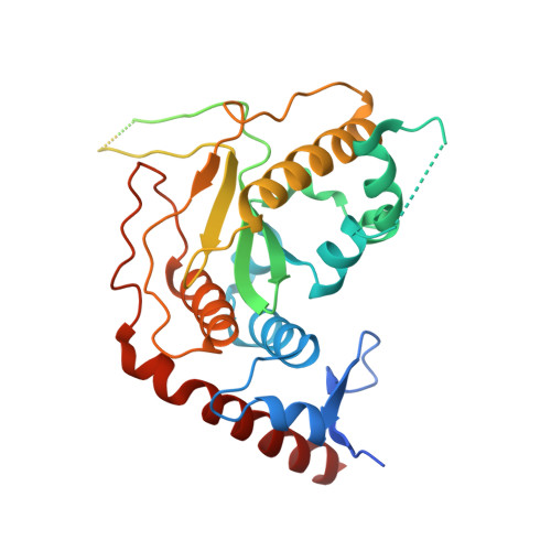
Structure of the Human SENP7 Catalytic Domain and Poly-SUMO Deconjugation Activities for SENP6 and SENP7.

Lima, C.D.Reverter, D.

(2008) J Biological Chem 283: 32045-32055

  • DOI: https://doi.org/10.1074/jbc.M805655200
  • Primary Citation Related Structures: 
    3EAY

  • PubMed Abstract: 

    Small ubiquitin-like modifier (SUMO) proteases regulate the abundance and lifetime of SUMO-conjugated substrates by antagonizing reactions catalyzed by SUMO-conjugating enzymes. Six SUMO proteases constitute the human SENP/ULP protease family (SENP1-3 and SENP5-7). SENP6 and SENP7 include the most divergent class of SUMO proteases, which also includes the yeast enzyme ULP2. We present the crystal structure of the SENP7 catalytic domain at a resolution of 2.4 angstroms. Comparison with structures of human SENP1 and SENP2 reveals unique elements that differ from previously characterized structures of SUMO-deconjugating enzymes. Biochemical assays show that SENP6 and SENP7 prefer SUMO2 or SUMO3 in deconjugation reactions with rates comparable with those catalyzed by SENP2, particularly during cleavage of di-SUMO2, di-SUMO3, and poly-SUMO chains composed of SUMO2 or SUMO3. In contrast, SENP6 and SENP7 exhibit lower rates for processing pre-SUMO1, pre-SUMO2, or pre-SUMO3 in comparison with SENP2. Structure-guided mutational analysis reveals elements unique to the SENP6 and SENP7 subclass of SENP/ULP proteases that contribute to protease function during deconjugation of poly-SUMO chains.


  • Organizational Affiliation
    • Structural Biology Program, Sloan-Kettering Institute, New York, New York 10065, USA. limac@mskcc.org

Macromolecule Content 

  • Total Structure Weight: 37.93 kDa 
  • Atom Count: 2,129 
  • Modeled Residue Count: 247 
  • Deposited Residue Count: 323 
  • Unique protein chains: 1

Macromolecules

Find similar proteins by:|  3D Structure
Entity ID: 1
MoleculeChains  Sequence LengthOrganismDetailsImage
Sentrin-specific protease 7323Homo sapiensMutation(s): 0 
Gene Names: SENP7KIAA1707SSP2SUSP2
EC: 3.4.22
UniProt & NIH Common Fund Data Resources
Find proteins for Q9BQF6 (Homo sapiens)
Explore Q9BQF6 
Go to UniProtKB:  Q9BQF6
PHAROS:  Q9BQF6
GTEx:  ENSG00000138468 
Entity Groups
Sequence Clusters30% Identity50% Identity70% Identity90% Identity95% Identity100% Identity
UniProt GroupQ9BQF6
Sequence Annotations
Expand
Reference Sequence

Small Molecules

Ligands 1 Unique
IDChains Name / Formula / InChI Key2D Diagram3D Interactions
SO4

Query on SO4


Download:Ideal Coordinates CCD File
B [auth A]SULFATE ION
O4 S
QAOWNCQODCNURD-UHFFFAOYSA-L

Experimental Data & Validation

Experimental Data

  • Method: X-RAY DIFFRACTION
  • Resolution: 2.40 Å
  • R-Value Free:  0.256 (Depositor), 0.194 (DCC) 
  • R-Value Work:  0.202 (Depositor), 0.204 (DCC) 
Space Group: P 32 2 1
Unit Cell:
Length ( Å )Angle ( ˚ )
a = 76.22α = 90
b = 76.22β = 90
c = 103.46γ = 120
Software Package:
Software NamePurpose
DENZOdata reduction
SCALEPACKdata scaling
SOLVEphasing
RESOLVEphasing
CNSrefinement
PDB_EXTRACTdata extraction
ADSCdata collection

Structure Validation

View Full Validation Report


Entry History 

Deposition Data

Revision History  (Full details and data files)

  • Version 1.0: 2008-09-16
    Type: Initial release
  • Version 1.1: 2011-07-13
    Changes: Version format compliance
  • Version 1.2: 2017-10-25
    Changes: Refinement description
  • Version 1.3: 2024-02-21
    Changes: Data collection, Database references, Derived calculations