3E86 | pdb_00003e86

High resolution Crystal Structure of the open NaK channel pore


Experimental Data Snapshot

  • Method: X-RAY DIFFRACTION
  • Resolution: 1.60 Å
  • R-Value Free: 
    0.242 (Depositor), 0.217 (DCC) 
  • R-Value Work: 
    0.212 (Depositor), 0.212 (DCC) 

wwPDB Validation 3D Report Full Report

Validation slider image for 3E86

This is version 1.4 of the entry. See complete history

Literature

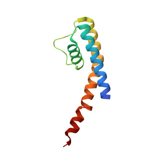
High-resolution structure of the open NaK channel

Alam, A.Jiang, Y.

(2009) Nat Struct Mol Biol 16: 30-34

  • DOI: https://doi.org/10.1038/nsmb.1531
  • Primary Citation Related Structures: 
    3E86

  • PubMed Abstract: 

    We report the crystal structure of the nonselective cation channel NaK from Bacillus cereus at a resolution of 1.6 A. The structure reveals the intracellular gate in an open state, as opposed to the closed form reported previously, making NaK the only channel for which the three-dimensional structures of both conformations are known. Channel opening follows a conserved mechanism of inner helix bending using a flexible glycine residue, the gating hinge, seen in MthK and most other tetrameric cation channels. Additionally, distinct inter and intrasubunit rearrangements involved in channel gating are seen and characterized for the first time along with inner helix twisting motions. Furthermore, we identify a residue deeper within the cavity of the channel pore, Phe92, which is likely to form a constriction point within the open pore, restricting ion flux through the channel. Mutating this residue to alanine causes a subsequent increase in ion-conduction rates as measured by (86)Rb flux assays. The structures of both the open and closed conformations of the NaK channel correlate well with those of equivalent K(+) channel conformations, namely MthK and KcsA, respectively.


  • Organizational Affiliation
    • Department of Physiology, University of Texas Southwestern Medical Center, 5323 Harry Hines Boulevard, Dallas, Texas 75390-9040, USA.

Macromolecule Content 

  • Total Structure Weight: 22.05 kDa 
  • Atom Count: 1,636 
  • Modeled Residue Count: 187 
  • Deposited Residue Count: 192 
  • Unique protein chains: 1

Macromolecules

Find similar proteins by:|  3D Structure
Entity ID: 1
MoleculeChains  Sequence LengthOrganismDetailsImage
Potassium channel protein
A, B
96Bacillus cereusMutation(s): 0 
Membrane Entity: Yes 
UniProt
Find proteins for Q81HW2 (Bacillus cereus (strain ATCC 14579 / DSM 31 / CCUG 7414 / JCM 2152 / NBRC 15305 / NCIMB 9373 / NCTC 2599 / NRRL B-3711))
Explore Q81HW2 
Go to UniProtKB:  Q81HW2
Entity Groups
Sequence Clusters30% Identity50% Identity70% Identity90% Identity95% Identity100% Identity
UniProt GroupQ81HW2
Sequence Annotations
Expand
Reference Sequence

Small Molecules

Ligands 4 Unique
IDChains Name / Formula / InChI Key2D Diagram3D Interactions
CS

Query on CS


Download:Ideal Coordinates CCD File
D [auth A],
H [auth B]
CESIUM ION
Cs
NCMHKCKGHRPLCM-UHFFFAOYSA-N
MPD

Query on MPD


Download:Ideal Coordinates CCD File
C [auth A],
I [auth B]
(4S)-2-METHYL-2,4-PENTANEDIOL
C6 H14 O2
SVTBMSDMJJWYQN-YFKPBYRVSA-N
CA

Query on CA


Download:Ideal Coordinates CCD File
E [auth A]CALCIUM ION
Ca
BHPQYMZQTOCNFJ-UHFFFAOYSA-N
NA

Query on NA


Download:Ideal Coordinates CCD File
F [auth A],
G [auth A],
J [auth B],
K [auth B]
SODIUM ION
Na
FKNQFGJONOIPTF-UHFFFAOYSA-N

Experimental Data & Validation

Experimental Data

  • Method: X-RAY DIFFRACTION
  • Resolution: 1.60 Å
  • R-Value Free:  0.242 (Depositor), 0.217 (DCC) 
  • R-Value Work:  0.212 (Depositor), 0.212 (DCC) 
Space Group: I 4
Unit Cell:
Length ( Å )Angle ( ˚ )
a = 68.03α = 90
b = 68.03β = 90
c = 89.264γ = 90
Software Package:
Software NamePurpose
DENZOdata reduction
SCALEPACKdata scaling
MOLREPphasing
CNSrefinement
PDB_EXTRACTdata extraction
HKL-2000data reduction

Structure Validation

View Full Validation Report


Entry History 

Deposition Data

Revision History  (Full details and data files)

  • Version 1.0: 2008-12-23
    Type: Initial release
  • Version 1.1: 2011-07-13
    Changes: Version format compliance
  • Version 1.2: 2011-11-16
    Changes: Atomic model
  • Version 1.3: 2017-10-25
    Changes: Refinement description
  • Version 1.4: 2024-02-21
    Changes: Data collection, Database references, Derived calculations